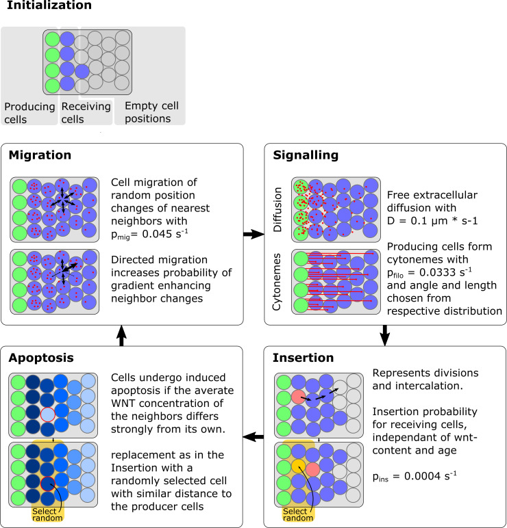
IMAGE

Fig 8

ID
ZDB-IMAGE-200720-9
Source
Figures for Rosenbauer et al., 2020
Image
Figure Caption

Fig 8 Overview over the simulation method and model parameters.

The field in which the tissue is modeled consists of precomputed non-overlapping potential cell positions (“fixed irregular lattice sites”). To initialize a simulation one margin of the field is filled with non-dividing morphogen producing cells (here shown in green), as well as morphogen receiving cells (shown in blue) with no initial morphogen concentration. After initialization (up to) four processes are sequentially executed for one timestep. In the signaling step morphogen is deposited from the producing into the receiving cells, either by diffusive transport or by direct cell to cell contacts via cytonemes, also some morphogen decays here in the recipient cells. Insertion drives the tissue growth and consists of two causes which cause the same effect, namely cell division and intercalation from overlying and underlying cell sheets. Here, if an insertion occurs at one cell, a path to the nearest empty cell position is determined and all cells are subsequently moved along this path, the emerging empty spot is than filled by a cell with its properties chosen randomly from all cells with similar distance to the producing cells. Apoptosis can be induced if the Wnt concentration of a single cell differs strongly from its neighbors, this process is only activated in the last third of the simulation and only the 130 cells with the strongest discrepancy of Wnt contents to neighbors are removed and replaced similarly to insertion. Migration models nearest neighbor interactions by a random swapping of positions of nearest neighbor cells. Directed migration can be introduced if the swapping probabilities are changed so that gradient enhancing swaps become more likely.

Acknowledgments
This image is the copyrighted work of the attributed author or publisher, and ZFIN has permission only to display this image to its users. Additional permissions should be obtained from the applicable author or publisher of the image. Full text @ PLoS Comput. Biol.