IMAGE

Figure 9

ID
ZDB-IMAGE-200716-62
Source
Figures for Keller et al., 2020
Image
Figure Caption

Figure 9

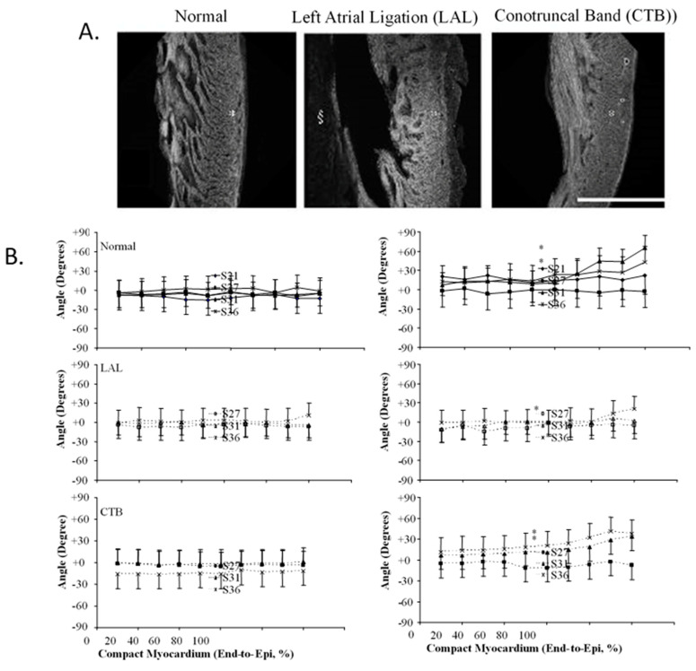
Three-dimensional myofiber architecture of the embryonic LV during normal development and altered mechanical loads. (A) Developmental change of the LV myocardial architecture in normal, left-atrial ligated (LAL), and conotruncal banded (CTB) embryos at stage 36. Scale bar = 500 μm. (B) Transmural myofiber angle distribution of the LV compact myocardium in normal, LAL, and CTB embryos. Data expressed as mean ± SD. Horizontal axis represents transmural coordinate of compact myocardium from endocardium (0%) to epicardium (100%). Asterisk, p < 0.05 by nonparametric ranking test vs. normal at the same developmental stage. This was adapted with permission [135].

Acknowledgments
This image is the copyrighted work of the attributed author or publisher, and ZFIN has permission only to display this image to its users. Additional permissions should be obtained from the applicable author or publisher of the image. Full text @ J Cardiovasc Dev Dis