
|
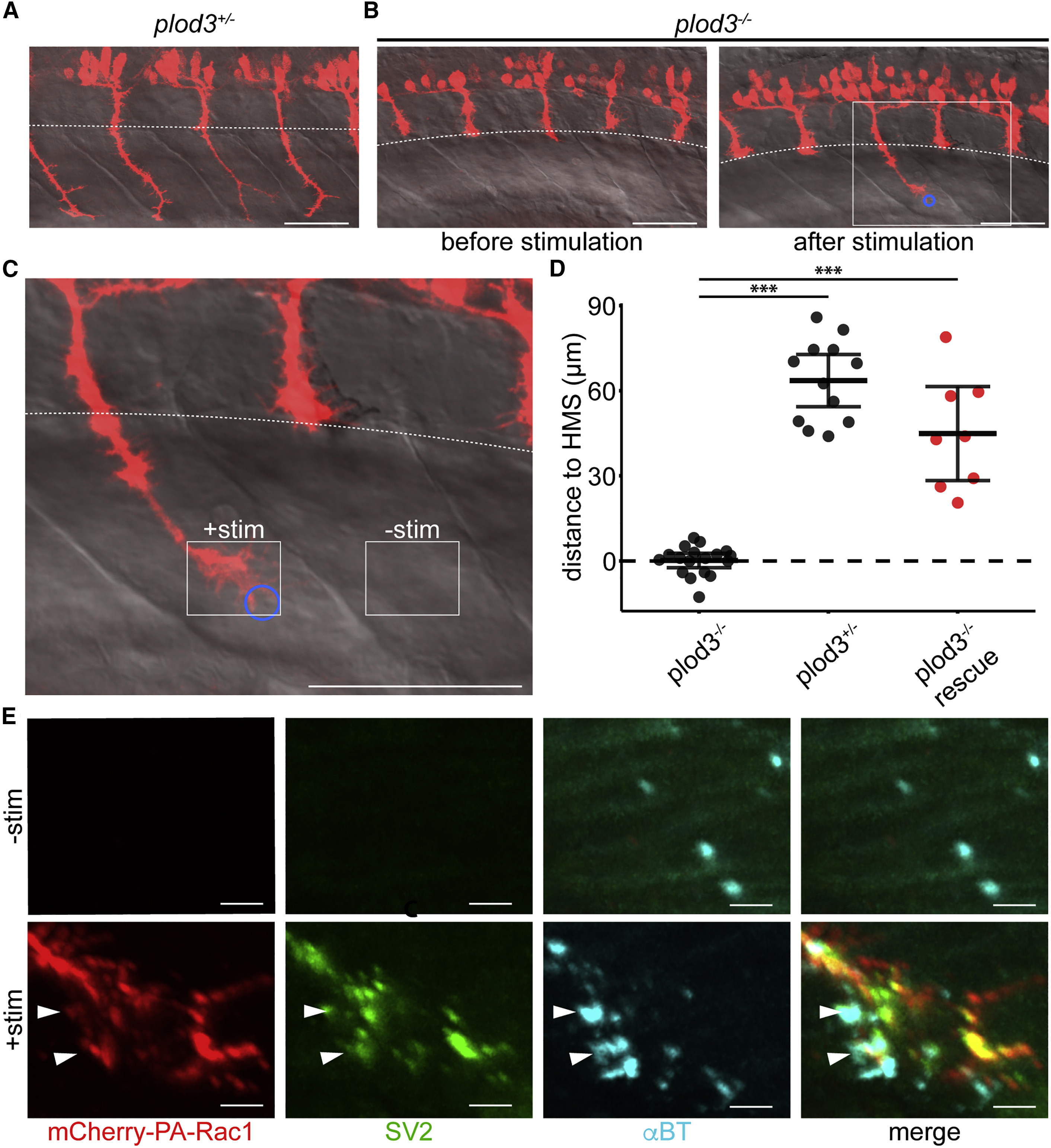
Fig. 4 Optogenetic Stimulation of plod3−/− Zebrafish CaP Neurons Rescues Their Axon Guidance Defect, Allowing Juxtaposition of Pre- and Post-synaptic Machinery within the Ventral Myotome (A) PA-Rac1 expression (red) in spinal motor neurons of plod3+/− zebrafish at 28 hpf (scale bar, 50 μm). (B) Left: an age-matched plod3−/− mutant sibling prior to stimulation fails to extend CaP axons into the ventral myotome past the horizontal myoseptum (dashed white line). Right: In plod3−/− fish, a single PA-Rac1+ CaP axon extended into the ventral myotome after illumination, while unilluminated axons remain arrested at the horizontal myoseptum (blue circle, region of illumination; scale bar, 50 μm). (C) Enlarged image of plod3−/− CaP axon following stimulation (solid white box in B; scale bar, 50 μm). (D) Illumination of PA-Rac1+ CaP axons in plod3−/− mutant fish induced growth significantly farther past the horizontal myoseptum than unilluminated axons. There was no significant difference in the distance grown past the horizontal myoseptum in illuminated plod3−/− fish compared with plod+/− fish (FDR adjusted, difference of least square means from mixed linear model, n = 8 axons, one axon per fish, ∗∗∗p < 0.001, mean and 95% CIs shown). (E) Immunohistochemistry for mCherry (red, left), SV2 (green, middle left), and αBT (cyan, middle right) showing colocalization of pre- and post-synaptic markers (merge, right) in the ventral myotome of the stimulated axon (bottom), but not in the unstimulated axon (top; scale bar, 5 μm). Stimulated and unstimulated regions correspond to solid white boxes in (C). See also Video S4.
Reprinted from Developmental Cell, 53, Harris, J.M., Wang, A.Y., Boulanger-Weill, J., Santoriello, C., Foianini, S., Lichtman, J.W., Zon, L.I., Arlotta, P., Long-Range Optogenetic Control of Axon Guidance Overcomes Developmental Boundaries and Defects, 577-588.e7, Copyright (2020) with permission from Elsevier. Full text @ Dev. Cell