IMAGE

Fig. 8

ID
ZDB-IMAGE-200629-24
Antibodies
Source
Figures for Chen et al., 2020
Image
Figure Caption

Fig. 8

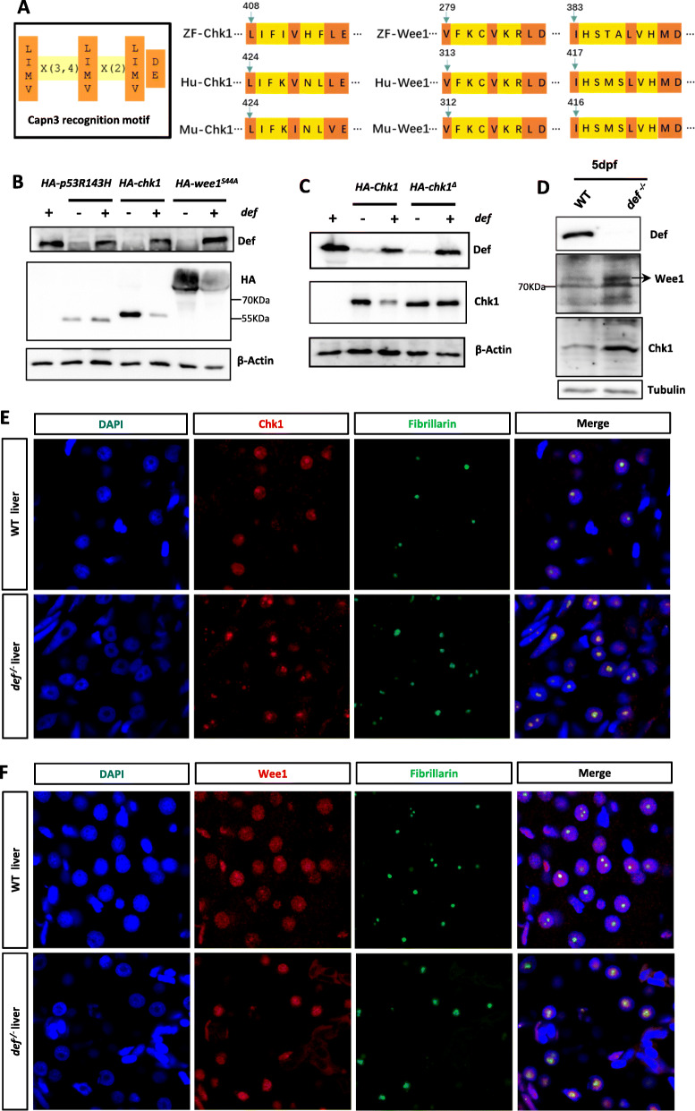
Chk1 and Wee1 are substrates of the Def-Capn3b complex. a Diagram showing the Capn3 recognition motif (left panel) and the locations of this motif in zebrafish, human and mouse Chk1 (middle panel) and Wee1 (right panel) proteins. b, c Western blot of Def, HA-p53R143H, HA-Chk1 and HA-Wee1S44A in WT embryos injected with HA-p53R143H, HA-Chk1 or HA-Wee1S44A mRNA combined with or without def mRNA (b). Western blot of HA-Chk1 and HA-Chk1Δ in WT embryos injected with HA-Chek1or HA-Chek1Δ mRNA combined with or without def mRNA (c). Total proteins were extracted at 8-h post-injection. β-Actin: loading control. d Western blot of the endogenous Chk1 and Wee1 proteins in WT and def−/− mutant embryos at 5dpf. Tubulin: loading control. e, f Co-immunostaining of Chk1 (e) or Wee1 (F) with Fibrillarin in the liver of WT with def−/− at 5dpf. DAPI: stain nuclei

Figure Data
Acknowledgments
This image is the copyrighted work of the attributed author or publisher, and ZFIN has permission only to display this image to its users. Additional permissions should be obtained from the applicable author or publisher of the image. Full text @ Cell Regen (Lond)