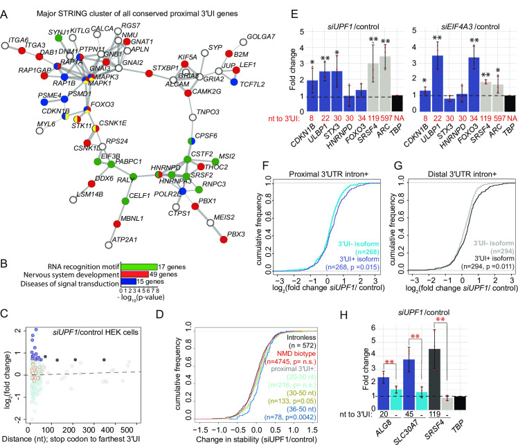
IMAGE

Fig 7

ID
ZDB-IMAGE-200629-1
Source
Figures for Gangras et al., 2020
Image
Figure Caption

Fig 7 Proximal position of 3′UTR introns is conserved in many vertebrate genes and a subset of proximal 3′UI-containing genes are NMD-sensitive in human cells.

A. A major interaction cluster predicted by STRING network analysis of genes with a 3′UI in proximal position in zebrafish, mouse and human. Nodes are colored by gene/protein function: nervous system (red), presence of RNA recognition motif (RRM) (green), diseases of signal transduction (blue), FoxO signaling pathway (yellow). (167 nodes and 127 edges in total, PPI enrichment p-value = 0.02). B. Gene ontology enrichment analysis of all 167 genes with conserved 3′UI proximal positioning. The most significant GO term within the following functional categories are shown: Interpro domains, Biological process and Reactome pathways. C. A scatter plot showing fold changes for APPRIS-annotated proximal 3′UI transcripts (dark blue: FC > 1.5 and light blue: FC < 1.5) and distal 3′UI transcripts (black: FC > 1.5 and gray: FC < 1.5) in UPF1 knockdown HEK293 cells compared to control cells [57]. Dots encircled in red are transcripts that also contain an uORF as determined previously in [57]. Same analysis for Ensembl transcript annotations is shown in Fig S5G. D. Empirical CDF plot showing change in mRNA stability for different classes of NMD targets and intron-less genes upon UPF1 knockdown in HEK293 cells (data from [57]). The gene classes are as follows: proximal 3′UI-containing genes where distance is 20–50 nts (light green), 30–50 nts (olive green) and 36–50 nts (dark blue), Ensembl-annotated NMD-biotype genes (red) and intron-less genes (black). CDF function is plotted as in Fig 5B. KS test p-value for comparison of NMD targets to intron-less genes is indicated in the same color. E. qRT-PCR analysis showing fold changes for proximal 3′UI-containing genes CDKN1B, FOXO3 (upregulated in zebrafish); ULBP1; STX3 (highest change in stability upon UPF1 KD in HEK293 cells [57]); HNRNPD (encodes RRM-containing protein) and distal 3′UI-containing genes (ARC and SRSF4) upon UPF1 (left) and EIF4A3 (right) knockdown in HCT116 cells. The distance between stop codon and 3′UI for every 3′UI-containing gene is indicated below each bar. TBP is the normalizing gene used for qRT-PCR analysis. Welch’s t-test p-values are indicated using asterisks (** p-value < 0.05 and * p-value < 0.1). F. Empirical CDF plot of fold changes in levels of 3′UI-containing isoforms (dark blue) as compared to 3′UI-lacking isoforms (sky blue) encoded from same genes in UPF1-depleted HEK293 cells. KS test p-value for differences in the two distributions is indicated on the bottom right. G. CDF plot as in F showing the fold changes in levels of distal 3′UI-containing (black) versus 3′UI-lacking isoforms (gray). H. qRT-PCR analysis showing fold changes for 3′UI-containing and 3′UI-lacking isoforms encoded by proximal 3′UI-containing genes ALG8 and SLC30A7 and by a distal 3′UI-containing gene SRSF4 upon UPF1 knockdown in HCT116 cells. The distance of 3′UI when present, or its absence (-) is indicated below each bar. TBP is the normalizing gene used for qRT-PCR analysis. Welch’s t-test p-values for the three biological replicates of each transcript are < 0.05. The Welch’s paired t-test p-values for the comparison between fold changes of 3′UI-containing and 3′UI-lacking isoforms is indicated using red asterisks (** p-value < 0.05).

Acknowledgments
This image is the copyrighted work of the attributed author or publisher, and ZFIN has permission only to display this image to its users. Additional permissions should be obtained from the applicable author or publisher of the image. Full text @ PLoS Genet.