
Fig. s3
- ID
- ZDB-IMAGE-200625-10
- Publication
- Weinschutz Mendes et al., 2020 - Expression of dlx genes in the normal and regenerating brain of adult zebrafish
- All Figures
- Figures for Weinschutz Mendes et al., 2020
|
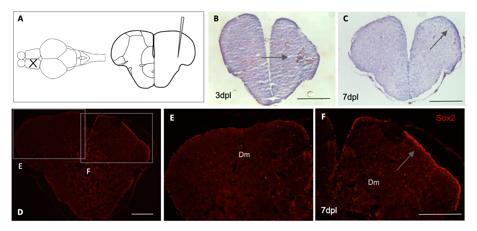
Fig. s3 Stab lesion and tissue integrity in adult zebrafish telencephalon. Schematic representation of lesion in the telencephalon area of adult zebrafish (A). Haematoxylin-Eosin staining and arrows indicate blood clot formation at 3 dpl (n = 3) (B) and apparent increase in cell number with higher intensity of staining at the lesioned hemisphere at 7dpl (n = 4) (C). Immunohistochemical labeling with Sox2 at 7 dpl indicates higher number of neural stem cells in the ventricular zone of the telencephalon (n = 3) (D). Magnification of image shown the contralateral hemisphere in E and regenerating hemisphere in F with increase in cells positive for Sox2 in the ventricular zone of the lesioned telencephalon. Scale bar (B-F): 200μM.