IMAGE

Fig 6

ID
ZDB-IMAGE-200620-14
Source
Figures for Ang et al., 2020
Image
Figure Caption

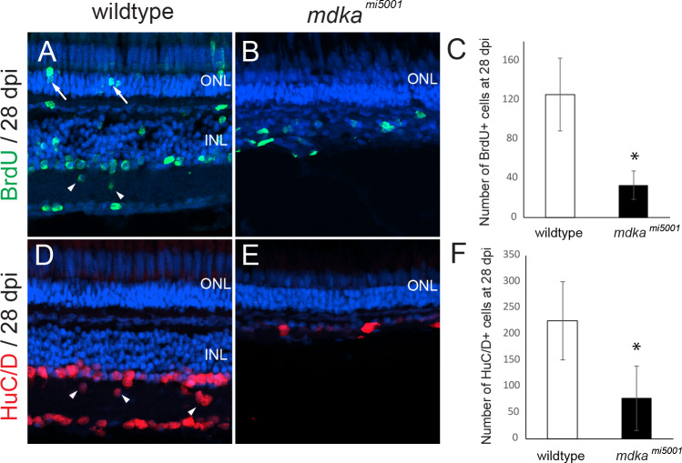
Fig 6 Absence of retinal regeneration in <italic>mdka</italic><sup><italic>mi5001</italic></sup> at 28 dpl following the death of inner retinal neurons.

(A, B) BrdU-labeled regenerated retinal neurons in wildtype (A) and mdkami5001 (B) retinas at 28 dpi. Arrows indicated BrdU-labeled rod photoreceptors. (C) The number of BrdU-positive cells wildtype (125.2 ± 37.1 cells) and mdkami5001 (33.2 ± 14.4 cells, p = 0.000341) retinas at 28 dpi. (D,E) Regenerated inner neurons labeled with HuC/D in wildtype (D) and mdkami5001 (E) at 28 dpi. Arrowheads indicate regenerated inner neurons are displaced in the inner plexiform layer. (F) The number of HuC/D cells in wildtype (224.8 ± 74.6 cells) and mdkami5001 (76.8 ± 61.4 cells, p = 0.00021) at 28 dpi. Student’s t-test, p*<0.05, n = 9 wildtype and 9 mutants. ONL: outer nuclear layer; INL: inner nuclear layer.

Acknowledgments
This image is the copyrighted work of the attributed author or publisher, and ZFIN has permission only to display this image to its users. Additional permissions should be obtained from the applicable author or publisher of the image. Full text @ PLoS One