IMAGE

Fig 3

ID
ZDB-IMAGE-200620-10
Source
Figures for Ang et al., 2020
Image
Figure Caption

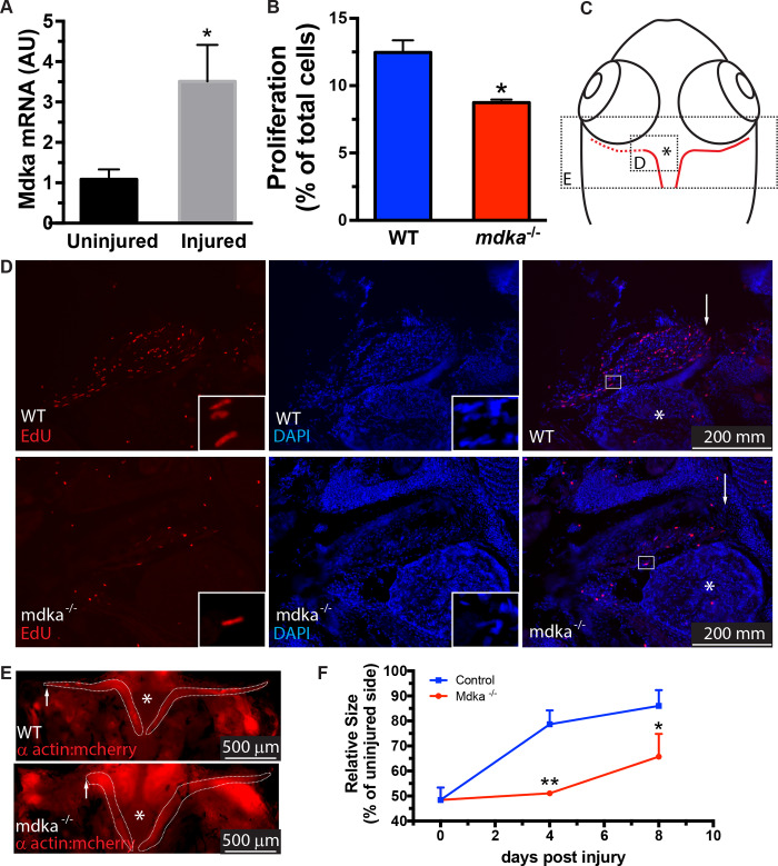
Fig 3 Regeneration of the extraocular muscle is delayed in <italic>mdka</italic><sup><italic>mi5001</italic></sup>.

(A) qPCR for midkine-a in wildtype zebrafish at 48 hours post lesion. Student’s t-test, p*<0.05, n = 3 uninjured and 3 injured (10 muscles per sample). (B) Cell counts of EdU labeled cells in muscles at 24 hours post lesion from wildtype (n = 4) and mdkami5001(n = 4). Student’s t-test, p*<0.05. (C) Diagram of the craniectomized head of an adult zebrafish. Red lines identify the lateral rectus muscles. Excised muscle is represented as red dashed line. Asterisk identifies the pituitary body at the midline. Boxes represent areas illustrated in panels D and E. (D) EdU-labeled cells in muscles from wildtype (top) and mdkami5001 (bottom) at 24 hours post lesion. High magnification inset image illustrates an elongated myonucleus undergoing proliferation (also see [13]). Arrows indicate the growing tip of the regenerating muscle. Asterisk indicates the pituitary body at the midline. (E) Extraocular muscles are outlined by dashed lines in control animals and in mutants at 4 days post lesion. Asterisks identify the pituitary body. (F) Outgrowth of regenerating muscles at post-injury time points in wildtype and mdkami5001. One-way ANOVA followed by Newman-Keuls multiple comparisons test, p*<0.05, p**<0.01, n = 4 wildtype and 4 mutants.

Acknowledgments
This image is the copyrighted work of the attributed author or publisher, and ZFIN has permission only to display this image to its users. Additional permissions should be obtained from the applicable author or publisher of the image. Full text @ PLoS One