IMAGE

Fig 4

ID
ZDB-IMAGE-200612-11
Source
Figures for Liu et al., 2020
Image
Figure Caption

Fig 4 Both TBC and rhodanese domains of TBC1D23 interact with golgin-97/245.

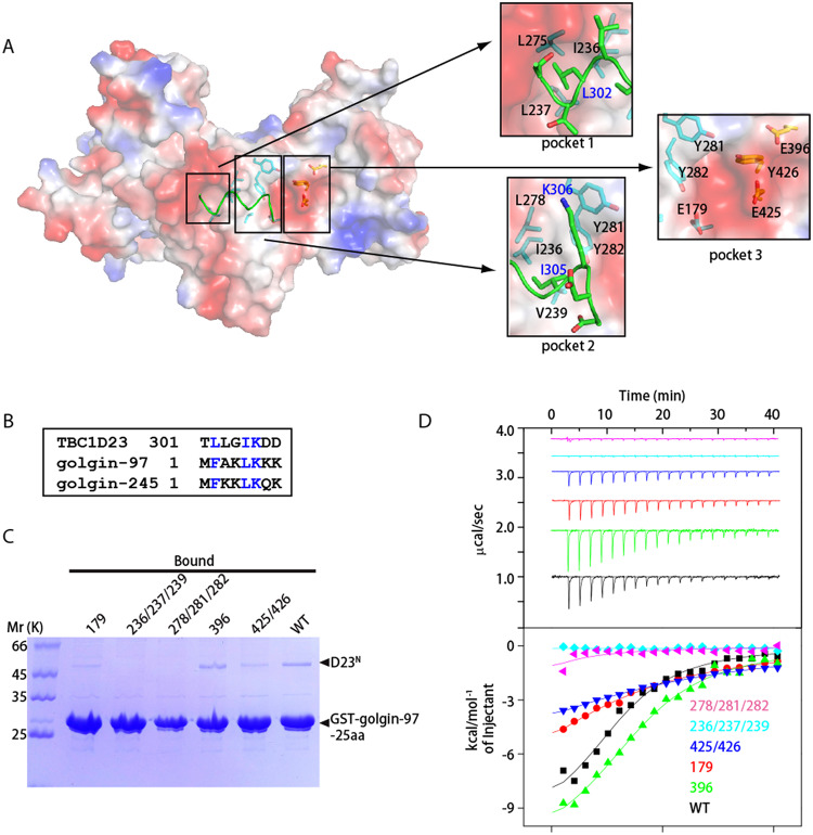
(A) Surface representation of one D23N monomer (molecule 1), with a fragment from symmetry-related D23N molecule shown as sticks (molecule 2, green). Molecule 1 is shown in the identical orientation to that in Fig 1B. Critical residues involved in dimerization are highlighted on the right, with residues from molecule 1 and 2 labeled in black and blue fonts, respectively. (B) Sequence alignment between residues from D23N-linker, N-terminus of golgin-97 and of golgin-245. Blue color indicates same or similar type of aa. (C) GST pull-down assays performed with GST-golgin-97-25aa and purified D23N WT, E179K (“179”), L278A/Y281A/Y282A (“278/281/282”), I236A/I237A/V239A (“236/237/239”), E396K (“396”), or E425K/Y426A (“425/426”). After incubation with soluble protein(s), the resin was extensively washed. The resin-bound proteins were then subjected to SDS-PAGE and Coomassie Blue staining. (D) ITC experiments for the binding of golgin-97-25aa peptide with D23N WT, E179K (“179”), L278A/Y281A/Y282A (“278/281/282”), I236A/I237A/V239A (“236/237/239”), E396K (“396”), or E425K/Y426A (“425/426”). Top and bottom panels show raw and integrated heat from injections, respectively. The solid curves in the bottom panel represent a fit of the integrated data to a single-site binding model. Experiments were triplicated, and the numerical data are included in S1 Data. aa, amino acid; D23N, N-terminus of TBC1D23; GST, glutathione S-transferase; ITC, isothermal titration calorimetry; TBC, Tre2-Bub2-Cdc16; WT, wild type.

Acknowledgments
This image is the copyrighted work of the attributed author or publisher, and ZFIN has permission only to display this image to its users. Additional permissions should be obtained from the applicable author or publisher of the image. Full text @ PLoS Biol.