IMAGE

Fig 5

ID
ZDB-IMAGE-200610-27
Source
Figures for Santos-Ledo et al., 2020
Image
Figure Caption

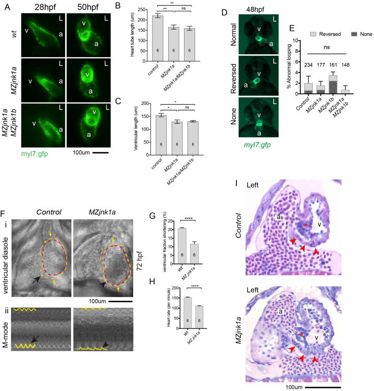
Fig 5 Cardiac abnormalities in <italic>MZjnk1a</italic> mutants.

(A) Live imaging of wildtype, MZjnk1a and MZjnk1a/ MZjnk1b mutants carrying myl7:gfp transgene at 28hpf and 50hpf. (B) overall primary heart tube length is reduced in MZjnk1b and MZjnk1a/ MZjnk1b embryos at 28hpf and (C) ventricular length is reduced at 50hpf. (D) Appearances of normal, reversed and no cardiac looping visualised by the myl7:gfp transgene in wildtype embryos. (E) Minimal levels of disordered cardiac looping in MZjnk1a and MZjnk1b mutants, alone and in combination. (F) Cardiac contractile function in MZjnk1a mutants and controls at 72 hpf. See S1 Movie, control, and S2 Movie, MZjnk1a mutant. External margin of the ventricle identified in diastole (yellow dashed line) and systole (red dashed line). i) Ventricular contraction is impaired in the FHF-derived ventricular segment (black arrowhead). ii) "M-mode" representations of the movies are obtained by resampling between the yellow arrows. Hypokinesis in the MZjnk1a heart (black arrowhead) can be seen, in comparison to the normal waveform of contraction in the control heart (black arrow). (G) Reduced fractional shortening and (H) heart rate in MZjnk1a mutants at 72 hpf. (I) Histological sections through heart of wildtype and MZjnk1a embryos in plane of imaging in (F). Red arrowheads indicate hypokinetic FHF ventricular segment, red asterix indicates atrioventricular valve. a = atrium, v = ventricle, L = left. * = p<0.05, ** = p<0.01, **** = p<0.0001, ns = not significant.

Figure Data
Acknowledgments
This image is the copyrighted work of the attributed author or publisher, and ZFIN has permission only to display this image to its users. Additional permissions should be obtained from the applicable author or publisher of the image. Full text @ PLoS Genet.