IMAGE

Figure 5

ID
ZDB-IMAGE-200608-7
Source
Figures for Albadri et al., 2020
Image
Figure Caption

Figure 5

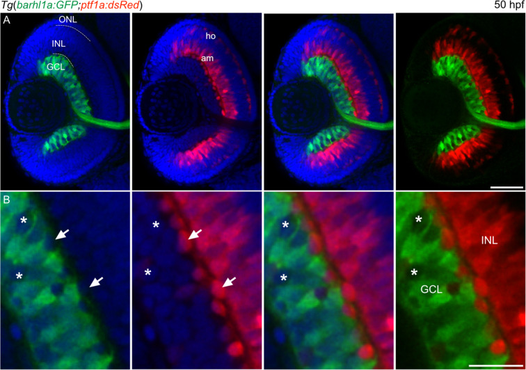
Barhl1a and Ptf1a reporter expressions are mutually exclusive. (A–B) Confocal imaging through the retina of a 50 hpf Tg(barhl1a:GFP;ptf1a:dsRed) embryo fixed and counterstained with DAPI (blue). Each image represents a single Z-stack in frontal view. Anterior is to the top. (B) always represents a higher magnification of (A). GFP-positive (green) cells are detected in the GCL whilst DsRed-positive cells (red) are detected in the amacrine (am) cells and horizontal (ho) cells of the inner nuclear layer (INL). White arrows point at DsRed-positive amacrine cells displaced in the GCL; which are always GFP-negative. Asterisks highlight the GFP-negative cells in the GCL; which are also DsRed-negative (B). GCL, ganglion cell layer; INL, inner nuclear layer; ONL, outer nuclear layer; am, amacrine cells; ho, horizontal cells. Scale bars: (A) 50 µm, (B) 20 µm.

Acknowledgments
This image is the copyrighted work of the attributed author or publisher, and ZFIN has permission only to display this image to its users. Additional permissions should be obtained from the applicable author or publisher of the image. Full text @ Sci. Rep.