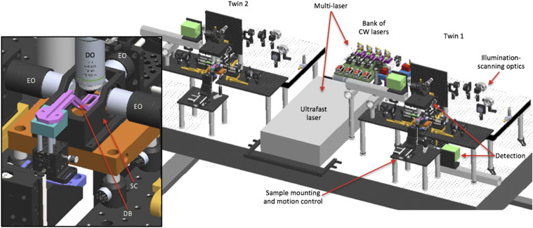
IMAGE

FIG. 2.

ID
ZDB-IMAGE-200606-3
Source
Figures for Keomanee-Dizon et al., 2020
Image
Figure Caption

FIG. 2.

3D opto-mechanical model of the twin-microscope system mounted on a 5 × 10 ft2, anti-vibration optical table. Model shows the multi-laser subsystem shared between microscope-twin-1 (right) and microscope-twin-2 (left). Twin-1 has the four functional subsystems labeled and features an implementation of both upright and inverted detection. Brief descriptions of each functional subsystem are provided in Table I. The inset shows a detailed view of the sample chamber (SC), the dive bar (DB) used to hold the sample, three excitation objectives (EO) to deliver excitation light-sheets to the sample, and the detection objective (DO) to collect emitted fluorescence from the sample.

Acknowledgments
This image is the copyrighted work of the attributed author or publisher, and ZFIN has permission only to display this image to its users. Additional permissions should be obtained from the applicable author or publisher of the image. Full text @ Rev. Sci. Instrum.