
Image
|
Figure Caption
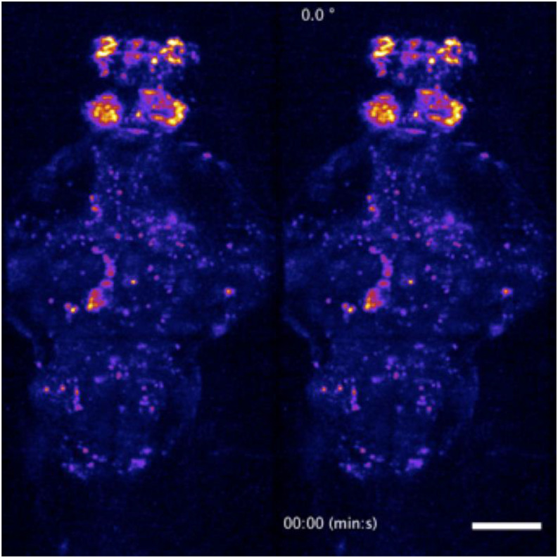
FIG. 13.
Dorsoventral (left) and rotating (right) maximum-intensity projections of a time-lapse recording of the whole-brain of the a 5-dpf transgenic larval zebrafish. Two-photon whole-brain functional light-sheet imaging was performed at a volumetric rate of 0.5 Hz. The video loops a 5-min recording as part of the data presented in
Acknowledgments
This image is the copyrighted work of the attributed author or publisher, and
ZFIN has permission only to display this image to its users.
Additional permissions should be obtained from the applicable author or publisher of the image.
Full text @ Rev. Sci. Instrum.