
Image
|
Figure Caption
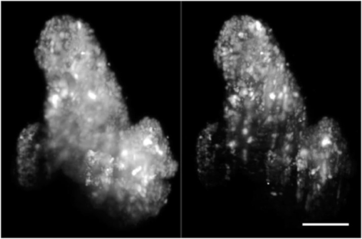
FIG. 11.
Volume rendering of fixed patient-derived tumor organoids expressing H2B-GFP, comparing images taken with one-photon (1P-, left) and two-photon excitation SPIM (2P-, right). Volumes are rotated around the
Acknowledgments
This image is the copyrighted work of the attributed author or publisher, and
ZFIN has permission only to display this image to its users.
Additional permissions should be obtained from the applicable author or publisher of the image.
Full text @ Rev. Sci. Instrum.