
|
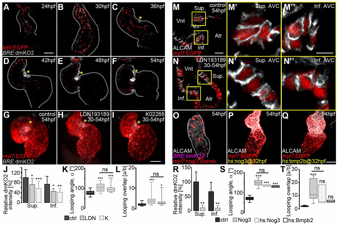
Fig. 3 Bmp signaling activity is asymmetric during S-looping morphogenesis. (A-F) Reconstructions of confocal z-scans of wild-type hearts expressing the Bmp signaling reporter transgene Tg(BRE:dmKO2)mw40 (false-colored gray) in combination with the Tg(kdrl:EGFP)s843 endocardial/endothelial reporter line (false-colored red) during heart looping morphogenesis (myocardium, dotted lines). (A,B) Initially, Bmp signaling is present within the entire endocardium and only weakly expressed within the myocardium. During early (C,D) and advanced (E,F) S-looping stages, BMP activity increases at the superior AVC (yellow asterisks) and more weakly at the inferior AVC. (G-L) Pharmacological inhibition of Bmp signaling using LDN193189 (20 μM) and K02288 (20 μM) inhibitors between 30 and 54 hpf. (G-I) Reconstruction of confocal z-stacks of the Bmp Tg(BRE:dmKO2)mw40 and myocardial Tg(mly7:EGFP)twu34 (false-colored red) reporter lines treated with (G) 0.2% DMSO (control), (H) LDN193189 or (I) K02288. Both LDN193189 and K02288 result in defective cardiac looping. Yellow asterisks highlight Bmp signaling within myocardium. (J-L) Bmp inhibition decreases the relative dmKO2 intensity at the superior (Sup.) and inferior (Inf.) AVC (J), increases the looping angle (α) (K) and decreases the looping overlap (increase in the index a/b) (L). (M-N″) Myocardial cell morphologies within the superior and inferior AVC after Bmp inhibition. (M,N) Single z-stack section images of ALCAM immunolabeled (false-colored white) and Tg(myl7:EGFP)twu34 myocardial reporter-expressing hearts (false-colored red) of embryos treated (M) with 0.2% DMSO (control) or (N) with LDN193189 between 30 and 54 hpf. (M′-N″) Enlargements of the areas outlined in M and N. Whereas myocardial cells at the superior AVC of control hearts have a columnar and conical shape (M′), the morphologies of AVC cells are cuboidal after Bmp inhibition (N′). Myocardial cells at the inferior AVC are cuboidal in control (M″) and in LDN-treated (N″) hearts. (O-T) Alterations in BMP signaling affect cardiac S-looping. (O-Q) Reconstructions of confocal z-scans of representative hearts. (O) Clonal overexpression of the BMP antagonist Noggin 3 causes a downregulation of the Bmp reporter transgene Tg(BRE:dmKO2)mw40 within the heart and an abnormal morphology. Overexpression of either (P) Noggin 3 [Tg(hsp70I:Nog3)fr14 transgenic line] or (Q) Bmp2B [Tg(hsp70:Bmp2b)fr13 transgenic line] by heat shock at 32 hpf causes cardiac looping defects at 54 hpf. (R) Clonal overexpression of Noggin 3 decreases expression of the Bmp reporter transgene Tg(BRE:dmKO2)mw40 at the superior (Sup.) and inferior (Inf.) AVC (Table S5). (S,T) Misregulated Bmp signaling causes an increase in looping angles (α) (Table S1) (S) and in the index a/b (Table S1) (T). In K,L,S,T, the limits of the boxes indicate the range between the first quartile (25th percentile) and the third quartile (75th percentile). The line inside box indicate the median value. The error bars indicate the maximum and minimum values. Data are mean±s.d. in J,R; ns, not significant; *P≤0.05; **P≤0.01; ***P≤0.001. Scale bars: 50 μm in A-I,O-Q; 20 μm in M,N; 10 μm in M′-N″.