IMAGE

Fig. S1

ID
ZDB-IMAGE-200523-29
Source
Figures for Saito et al., 2010
Image
Figure Caption

Fig. S1

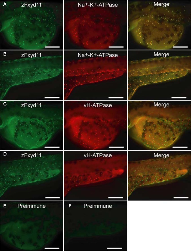
Localization of zFxyd11 in NaK-MRCs of the skin of zebrafish larvae. Immunofluorescence staining was performed on 2-dpf zebrafish larvae with anti-zFxyd11 antiserum [green in (A–D)], along with anti–Na+–K+-ATPase [red in (A), (B)] or anti–vH+-ATPase antiserum [red in (C), (D)]. Control staining was carried out with preimmune serum (E,F). Regions of the yolk sac (A,C,E) and the yolk sac extension and the trunk (B,D,F) are shown. Note that signals for zFxyd11 entirely overlap with NaK-MRCs on the skin of yolk, yolk extension, and trunk (A,B), and are not colocalized with vH-MRCs (C,D). Scale bars, 200 μm.

Acknowledgments
This image is the copyrighted work of the attributed author or publisher, and ZFIN has permission only to display this image to its users. Additional permissions should be obtained from the applicable author or publisher of the image. Full text @ Front. Physiol.