
Image
|
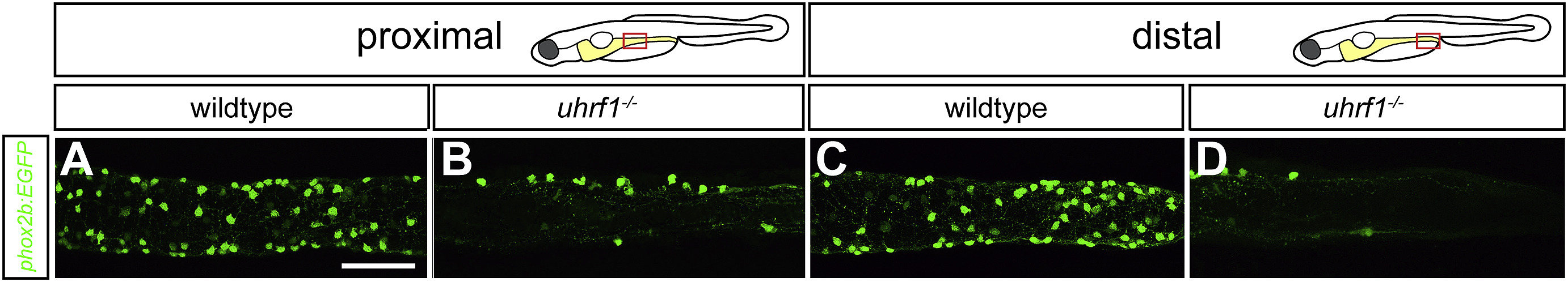
Figure Caption
Fig. 3 uhrf1 mutants have fewer neurons in both proximal and distal intestine. Confocal images of dissected intestines of wildtype (A,C) and uhrf1 mutants (B,D). uhfr1 mutants have fewer phox2b:EGFP positive enteric neurons (green) than wildtype siblings at 5 dpf (A,C) in both proximal (B) and distal (D) intestine. Scale bar = 50 μm.
Figure Data
Acknowledgments
This image is the copyrighted work of the attributed author or publisher, and
ZFIN has permission only to display this image to its users.
Additional permissions should be obtained from the applicable author or publisher of the image.
Reprinted from Developmental Biology, 455, Ganz, J., Melancon, E., Wilson, C., Amores, A., Batzel, P., Strader, M., Braasch, I., Diba, P., Kuhlman, J.A., Postlethwait, J.H., Eisen, J.S., Epigenetic factors Dnmt1 and Uhrf1 coordinate intestinal development, 473-484, Copyright (2019) with permission from Elsevier. Full text @ Dev. Biol.