
Image
|
Figure Caption
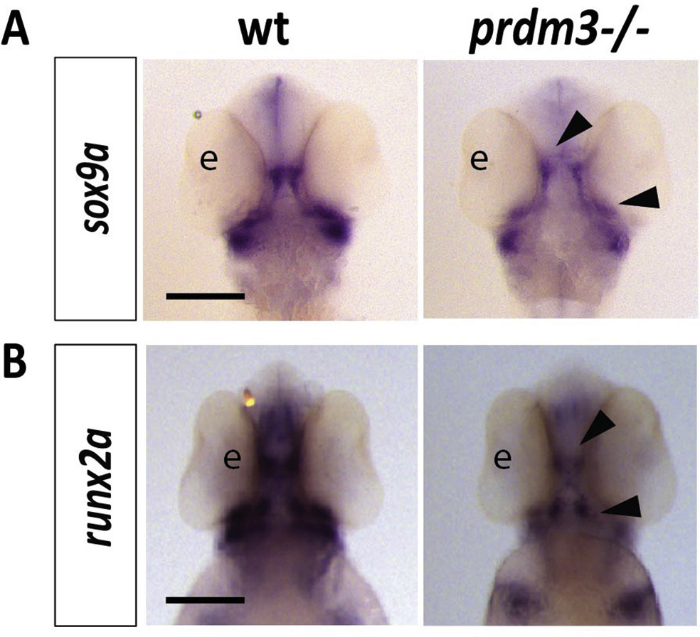
Fig. s2 Cartilage and bone markers, sox9a and runx2a, are reduced in the pharyngeal arches with loss of prdm3 in zebrafish. (A-B) Wildtype or prdm3−/− mutant embryos were collected and in situ hybridization was performed for sox9a (A) or runx2a (B) at 48 hpf. Shown are ventral views for each transcript. Black arrow heads indicate areas of decreased expression of these markers in the developing viscerocranium and neurocranium. Scale bars, 250 μm. Abbreviations: eye (e).
Acknowledgments
This image is the copyrighted work of the attributed author or publisher, and
ZFIN has permission only to display this image to its users.
Additional permissions should be obtained from the applicable author or publisher of the image.
Reprinted from Developmental Biology, 461(2), Shull, L.C., Sen, R., Menzel, J., Goyama, S., Kurokawa, M., Artinger, K.B., The conserved and divergent roles of Prdm3 and Prdm16 in zebrafish and mouse craniofacial development, 132-144, Copyright (2020) with permission from Elsevier. Full text @ Dev. Biol.