
|
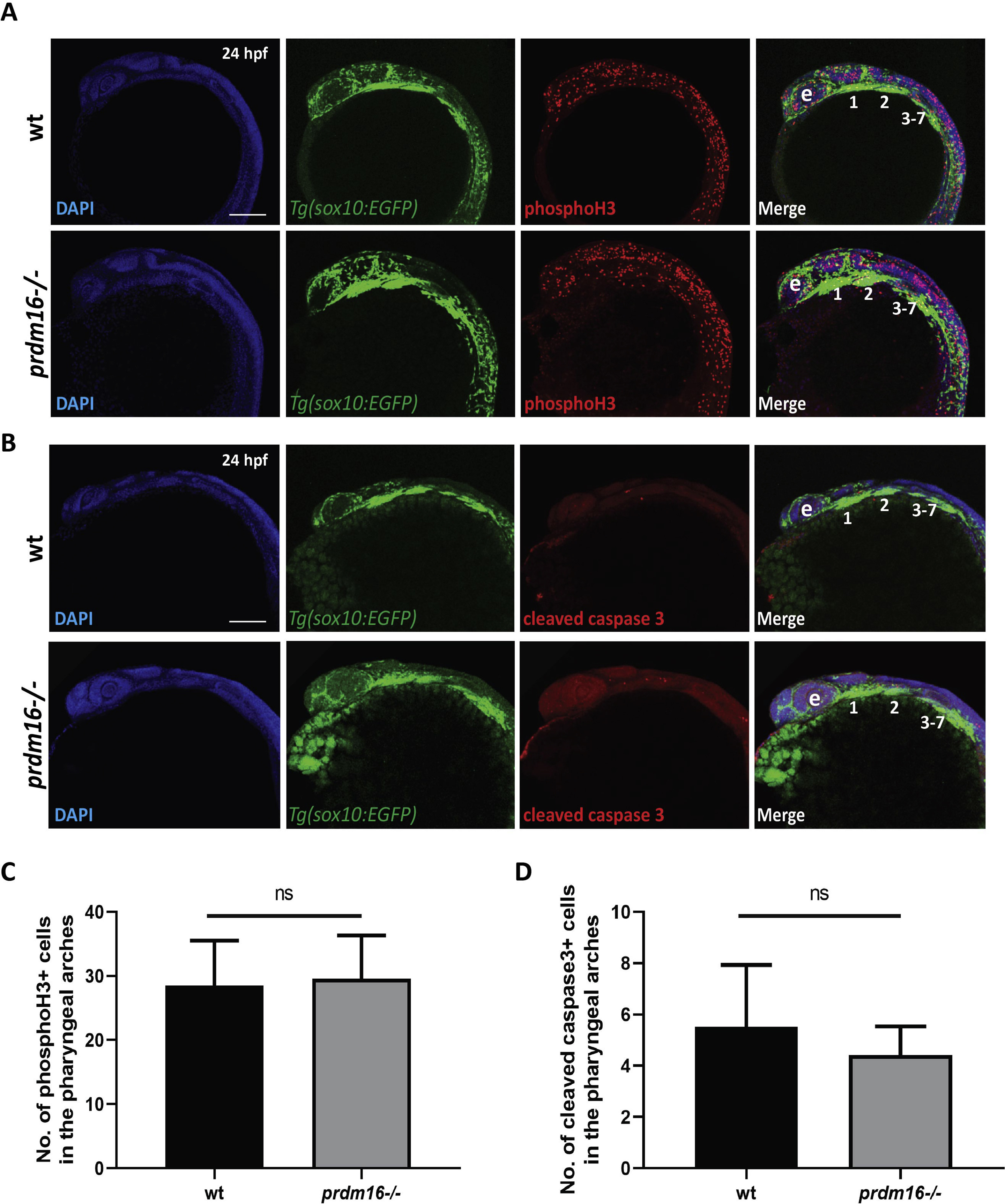
Fig. s7 Cell proliferation and cell death are not affected in the pharyngeal arches with loss of prdm16. (A-D) Wildtype or prdm16−/− zebrafish embryos crossed into the Tg(sox10:EGFP) transgenic line were collected at 24 hpf and whole mount immunofluorescence was performed for phosphorylated histone H3 (A) or cleaved caspase 3 (B) to assess cell proliferation or apoptosis in the pharyngeal arches, respectively. Shown are lateral max projection images of the pharyngeal arches 1–7. (C-D) Quantification of the number of phosphorylated histone H3 positive cells (yellow) (C) or cleaved caspase 3 positive cells (yellow) (D) within the pharyngeal arches (n = 6 embryos for each group). Abbreviations: eye (e), pharyngeal arches (1–7). Scale bar 100 μm ns, not significant, Student’s t-test.
Reprinted from Developmental Biology, 461(2), Shull, L.C., Sen, R., Menzel, J., Goyama, S., Kurokawa, M., Artinger, K.B., The conserved and divergent roles of Prdm3 and Prdm16 in zebrafish and mouse craniofacial development, 132-144, Copyright (2020) with permission from Elsevier. Full text @ Dev. Biol.