
Image
|
Figure Caption
Fig. 1
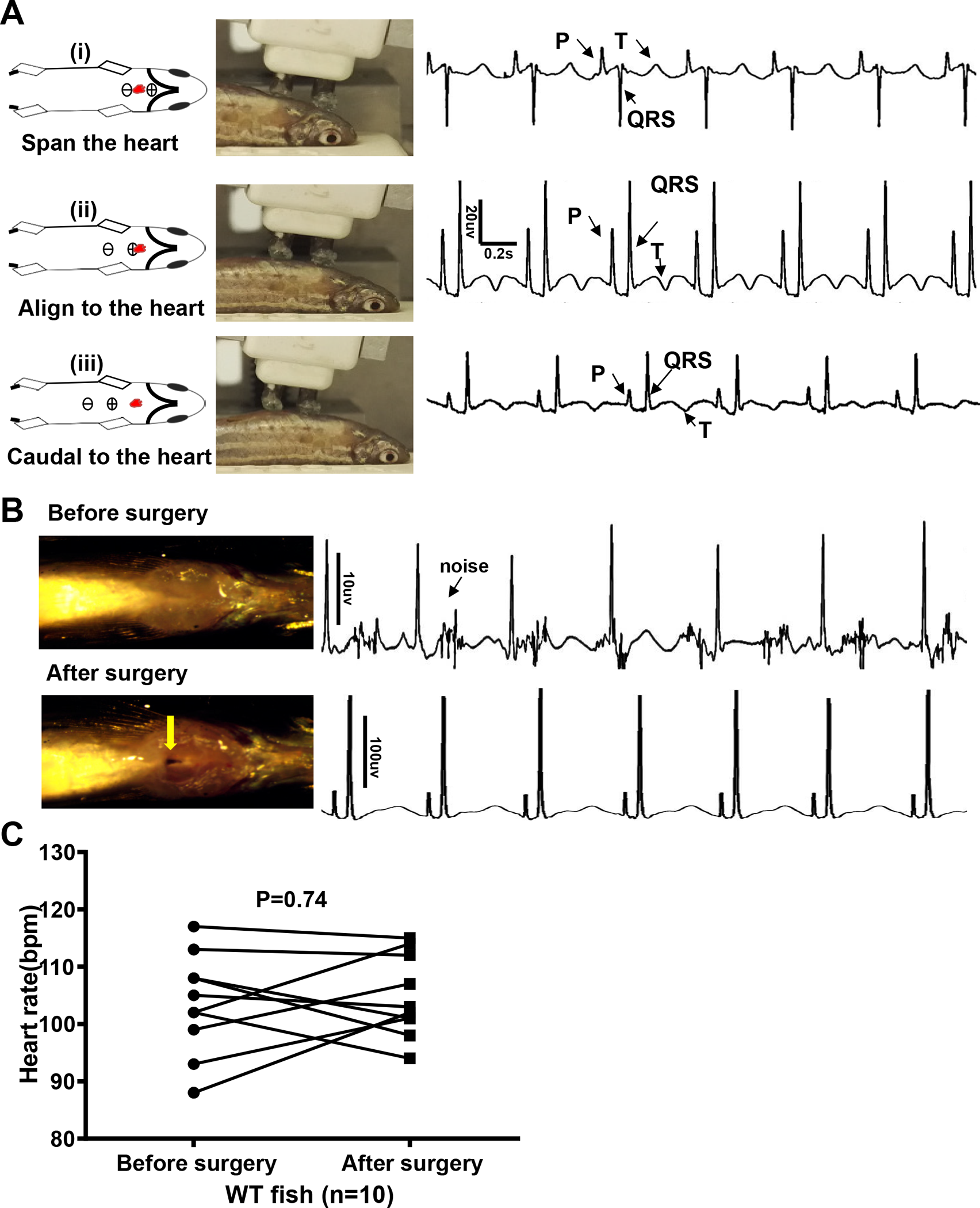
Quality of electrocardiography (ECG) signals is improved by optimizing location of the probes and microsurgery.
A, Different ECG waveform with distinct P, R, S, and T waves when probes were at a different location relative to the heart (red dots). We recommend aligning the positive probe to the heart (ii), whereby the ECG waveform is similar to that in humans. B, Microsurgery was conducted to open the pericardium sac and thus eliminate noise and enhance amplitude of the waveforms by about 10-fold. C, Change in heart rate with surgery. bpm indicates beats per minute; WT, wild-type.
Acknowledgments
This image is the copyrighted work of the attributed author or publisher, and
ZFIN has permission only to display this image to its users.
Additional permissions should be obtained from the applicable author or publisher of the image.
Full text @ PLoS One