
|
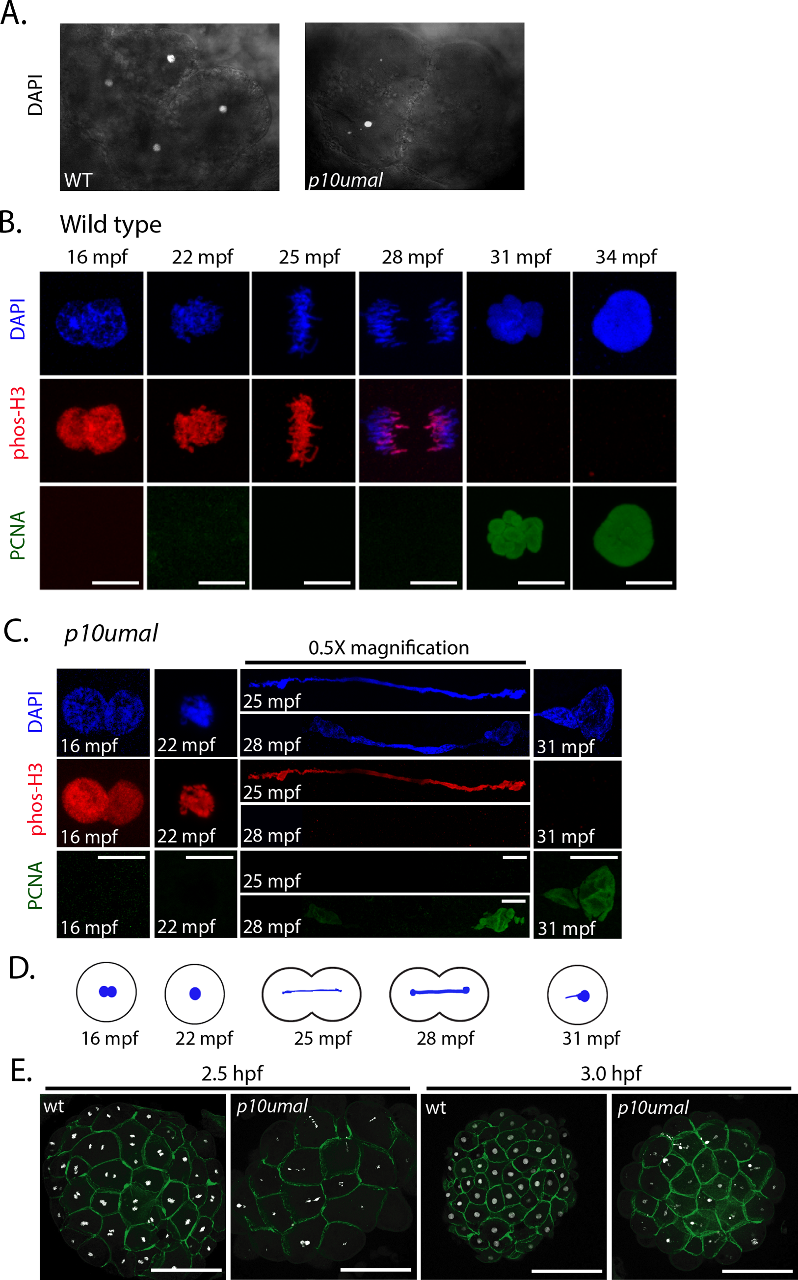
Fig. 3 Nuclear division is disrupted in p10umal mutants. A. DAPI staining of wild-type and p10umal 8-cell stage embryos (n = 11). Note: only four of the 8-cells are in view. B. Wild-type and C. p10umal fertilization time courses (N = 3 females examined). Embryos were fixed at 16, 22, 25, 28, 31 and 34 mpf. A minimum of five embryos corresponding to each time point were examined (representative images are shown). Pronuclei (16 mpf) and the one-cell zygote (at 22–34 mpf) were stained with DAPI (blue), anti-phospho-histone H3 (red), and anti-PCNA (green). Scale bars = 10μm. The 25 and 28 mpf time points were digitally reduced by 0.5x. D. Schematic representation of the p10umal phenotype at the corresponding time points illustrating the typical DNA bridge between dividing cells. E. Wild type and p10umal mutants at 2.5 and 3.0 hpf stained with DAPI and phalloidin to mark DNA and the cell boundaries, respectively. A minimum of 3 and up to 6 embryos each from 3 different females were examined for each time point (representative images are shown).