Mesoderm specification in blastomere reaggregates.
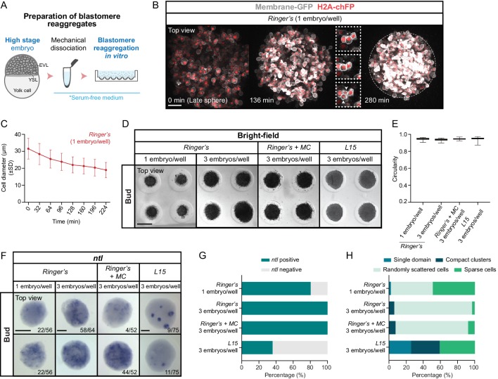
(A) Schematic representation of the preparation method of blastomere reaggregates from high stage embryos. (B) High-resolution fluorescence images of a blastomere reaggregate (top view) from late sphere stage onwards (30–45 min after reaggregate preparation; n = 8, N = 8). Cell membranes (grey) are marked by Membrane-RFP and nuclei (red) by H2A-chFP expression. Time in min. The reaggregate edges at the end of the acquisition are outlined by a white dashed line. Insets are zoom-in images of a cell division event completed at 136 min (dashed boxes). The dividing cell is highlighted with an asterisk and the two daughter cells with white arrowheads. (C) Cell diameter in blastomere reaggregates from late sphere stage onwards (0 min: number of cells = 211, n = 8, N = 8; 32 min: number of cells = 209, n = 8, N = 8; 64 min: number of cells = 208, n = 8, N = 8; 96 min: number of cells = 199, n = 8, N = 8; 128 min: number of cells = 197, n = 8, N = 8; 160 min: number of cells = 198, n = 8, N = 8; 192 min: number of cells = 210, n = 8, N = 8; 224 min: number of cells = 199, n = 8, N = 8). (D) Bright-field single-plane images of blastomere reaggregates (top view) prepared at a density of one or three embryos/well (indicated at the top) and subsequently cultured in either Ringer’s (one embryo/well: n = 233, N = 3; three embryos/well: n = 202, N = 3), Ringer’s with methylcellulose (MC 0.3%; three embryos/well: n = 184, N = 3) or L15 (three embryos/well: n = 227, N = 3) media until bud stage. (E) Circularity of bud stage blastomere reaggregates prepared at a density of one or three embryos/well (indicated at the side) and cultured in either Ringer’s (one embryo/well: n = 233, N = 3; three embryos/well: n = 202, N = 3), Ringer’s with methylcellulose (MC 0.3%; three embryos/well: n = 184, N = 3) or L15 (three embryos/well: n = 227, N = 3) media. (F) Expression of the mesodermal marker ntl, as determined by whole mount in situ hybridization in blastomere reaggregates (top view) prepared at a density of one or three embryos/well (indicated at the top) and subsequently cultured in either Ringer’s (one embryo/well: n = 56, N = 4; three embryos/well: n = 64, N = 4), Ringer’s with methylcellulose (MC 0.3%; three embryos/well: n = 52, N = 3) or L15 (three embryos/well: n = 75, N = 5) media until bud stage. The proportion of blastomere reaggregates with a phenotype similar to the images shown is indicated in the lower right corner. (G) Percentage of bud stage blastomere reaggregates prepared at a density of one or three embryos/well (indicated at the side) and cultured in either Ringer’s (one embryo/well: n = 56, N = 4; three embryos/well: n = 64, N = 4), Ringer’s with methylcellulose (MC 0.3%; three embryos/well: n = 52, N = 3) or L15 (three embryos/well: n = 75, N = 5) media showing or not ntl expression, as determined by whole mount in situ hybridization. (H) Percentage of bud stage blastomere reaggregates prepared at a density of one or three embryos/well (indicated at the side) and cultured in either Ringer’s (one embryo/well: n = 45, N = 4; three embryos/well: n = 64, N = 4), Ringer’s with methylcellulose (MC 0.3%; three embryos/well: n = 52, N = 3) or L15 (three embryos/well: n = 27, N = 4) media showing a single coherent ntl expression domain, several compact ntl-positive cell clusters (example shown for L15 cultured reaggregates in F), few sparse ntl positive cells (example shown for Ringer’s, Ringer’s + MC and L15 cultured reaggregates in F) or randomly scattered groups of ntl expressing cells (examples shown for Ringer’s or Ringer’s + MC cultured reaggregates in F), as determined by whole mount in situ hybridization. Scale bars: 100 µm (B), 500 µm (D), 150 µm (F).
Mesoderm specification in blastomere reaggregates.
Acknowledgments
This image is the copyrighted work of the attributed author or publisher, and
ZFIN has permission only to display this image to its users.
Additional permissions should be obtained from the applicable author or publisher of the image.
Full text @ Elife
Your Input Welcome
Thank you for submitting comments. Your input has been emailed to ZFIN curators who may contact you if
additional information is required.
Oops. Something went wrong. Please try again later.