
|
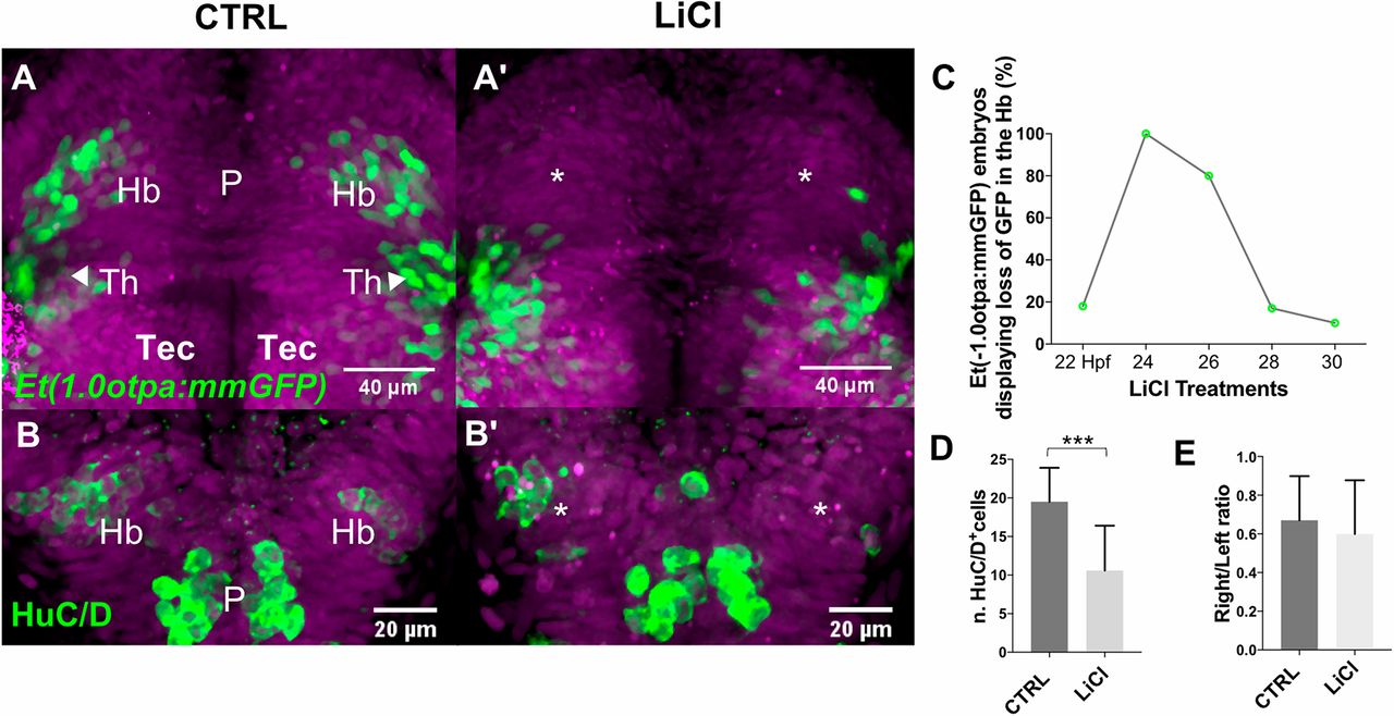
Fig. 2 Premature intrinsic activation of Wnt signaling delays habenular neuron differentiation. (A-B′) Projections of confocal z-stacks, dorsal views, anterior is towards the top focused onto the diencephalon of (A,A′) Et(-1.0otpa:mmGFP) and (B,B′) tg(flh:GFP); tg(foxD3:GFP) transgenic embryos. Nuclei are DAPI labeled (purple). (A,A′,C) Transient Wnt signaling activation causes a specific loss of GFP-expressing habenular neurons at 48 hpf. (B,B′,D,E) The number of HuC/D-positive differentiating habenular neurons is reduced at 36 hpf; their left-right asymmetric development remains unchanged. CTRL, control; Hb, habenulae; P, pineal; Tec, optic tectum; Th, thalamus.