IMAGE

Figure 3

ID
ZDB-IMAGE-200423-70
Source
Figures for Cozzolino et al., 2020
Image
Figure Caption

Figure 3

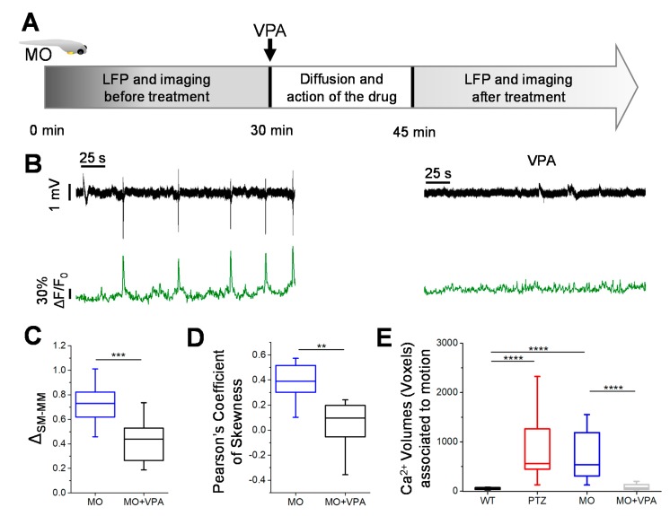
Valproate treatment. (A) Valproate (2 mM) was administered to MO kcnj10a larvae after 30 min of baseline recording of LFP and two-photon imaging. After a diffusion time of 15 min, the double recording was resumed. (B) Alignment of LFP and ΔF/F0 calcium traces in MO VPA( Valproic Acid)-treated larvae. (C) The analysis of the statistical distribution of the electrophysiological signal showed a clear effect of VPA in decreasing the energy of the LFP events. (D) The skewness of statistical distributions of Ca2+ fluctuations decreased during VPA treatment. (E) During VPA treatment, movements of the larvae did not correlate with a generalized increase of calcium signal. Therefore, VPA treatment led to a physiological phenotype, where tail flicks are associated not to a convulsive seizure but to the activation of a limited neuronal circuitry. 2 recordings for each larva, n = 3 larvae for each condition (* p ≤ 0.05, ** p ≤ 0.01, *** p ≤ 0.001, **** p ≤ 0.0001).

Figure Data
Acknowledgments
This image is the copyrighted work of the attributed author or publisher, and ZFIN has permission only to display this image to its users. Additional permissions should be obtained from the applicable author or publisher of the image. Full text @ Cells