IMAGE

Figure 1-S1

ID
ZDB-IMAGE-200423-110
Source
Figures for Lim et al., 2020
Image
Figure Caption

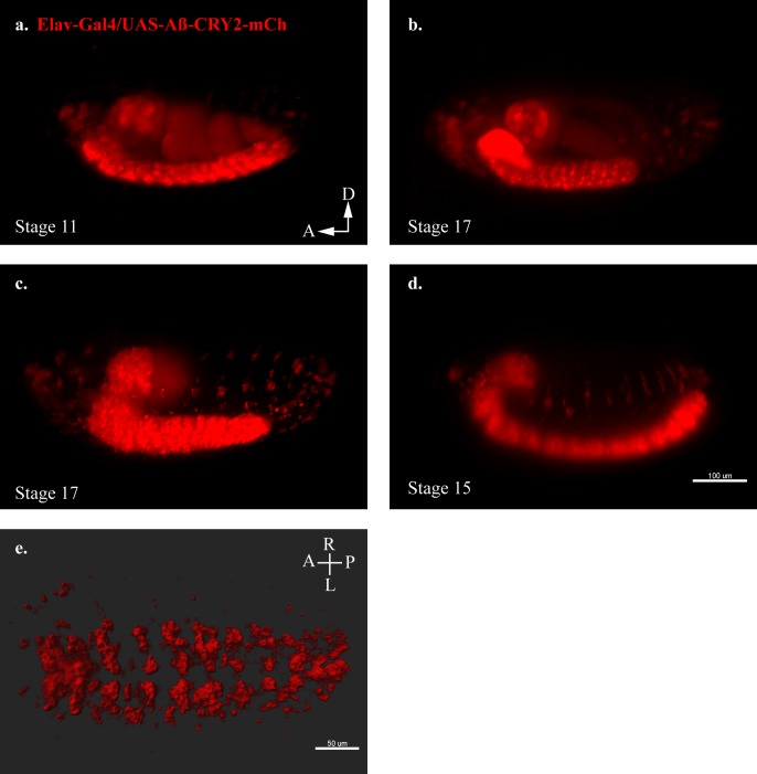
Figure 1-S1 Transgenic <italic>Drosophila</italic> model expressing Aβ-CRY2-mCh.

(a–d) Still images from lightsheet timelapse microscopy of Elav-Gal4/UAS-Aβ-CRY2-mCh in neurons detected by mCh fluorescence (red) in transgenic Drosophila embryos at different developmental stages. (e) Aβ localization to neuronal cell body volume in a Drosophila embryo expressing the mentioned construct.

Acknowledgments
This image is the copyrighted work of the attributed author or publisher, and ZFIN has permission only to display this image to its users. Additional permissions should be obtained from the applicable author or publisher of the image. Full text @ Elife