
|
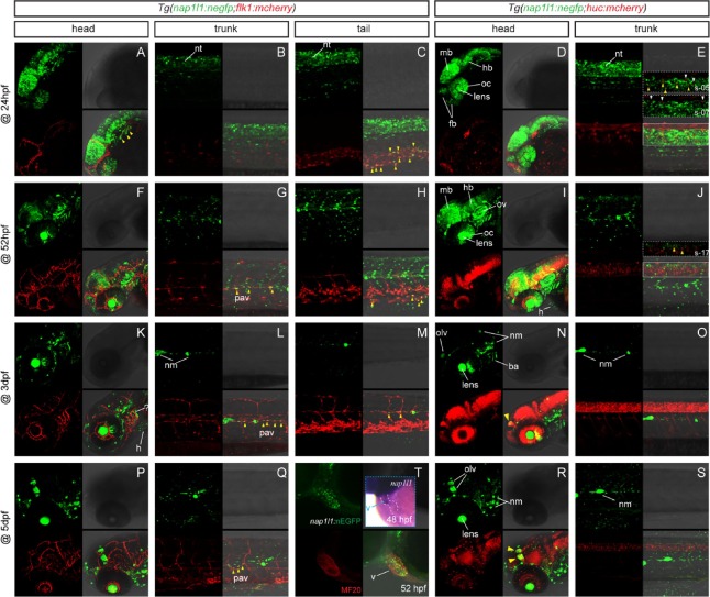
Fig. 2 Early expression of nap1l1 transgenic line. Confocal imaging for the developmentally staged embryos or larvae from the out-cross of Tg(nap1l1:nEGFP)zs102 with the endothelial reporter line Tg(flk1:mCherry) and the neuronal reporter line Tg(huc:mCherry), respectively. (T) Confocal immunofluorescent imaging of the 52-hpf Tg(nap1l1:nEGFP)zs102 embryo with primary antibody MF20 (red). Inset demonstrates nap1l1-expressing cells within the developing heart (ventricle), which was analyzed by whole-mount in situ hybridization with antisense probe of nap1l1 mRNA. Abbreviations: ba, branchial arches; fb, forebrain; h, heart; hb, hindbrain; mb, midbrain; nm, neuromast; nt, neural tube; oc, optic cup; olv, olfactory vesicle; ov, otic vesicle; pav, paraxial vessel; v, ventricle. (For interpretation of the references to colour in this figure legend, the reader is referred to the web version of this article.)
Reprinted from Gene, 735, Sun, S., Liu, Z., Li, X., Jia, J., Zhang, G., Yang, C., Jiang, Q., Zou, Y., Characterization of a nap1l1 transgenic reporter in zebrafish, 144388, Copyright (2020) with permission from Elsevier. Full text @ Gene