
|
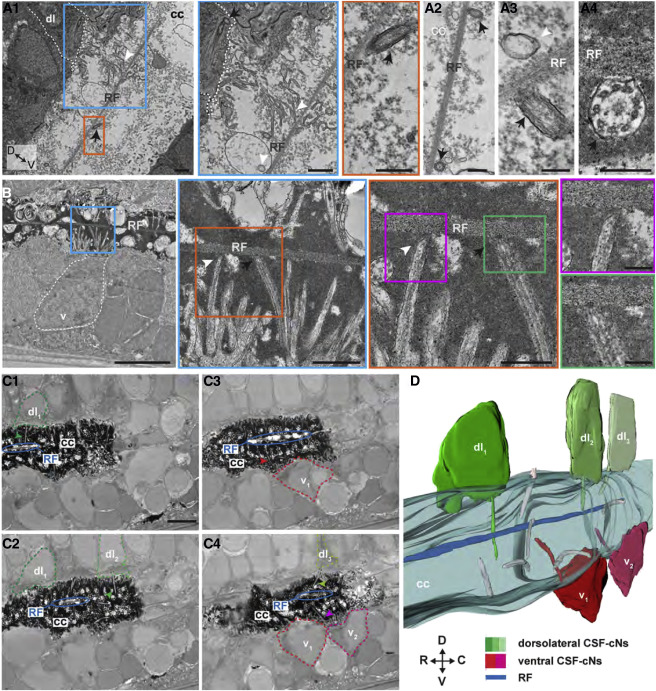
Fig. 6 The RF Can Contact Cilia and Microvilli of CSF-cNs in the Central Canal (A and B) Transmission electron microscopy (TEM) from spinal cord sectioned in the sagittal plane reveals motile cilium (black arrow) near or in contact with the RF as well as microvilli from CSF-cNs (white arrowhead) near RF. Zoomed regions highlighted in different colors. (A1) The RF tends to be dorsally located in the central canal and close to cilia (arrow), as well as microvilli (arrowhead) originating from identifiable dorsolateral CSF-cNs. (A2–A4) Appositions onto the RF of motile cilia, which contain two central microtubules along the axoneme (arrow in A2 and A4), as well as microvilli from a ventral CSF-cN (arrowhead, A3). (B) Zooms of lateral regions delineated in colored lines show that ventral CSF-cNs can apparently contact the RF via their microvilli (arrowheads) and their motile cilium (arrows). (C) Z projection stack of 3 subsequent images acquired in the sagittal plane from serial block face scanning electron microscopy (SBF-SEM), highlighting apparent contacts between RF (blue ellipse) and dorsolateral CSF-cNs. (C1)–(C4) correspond to different positions in the sagittal plane where multiple dorsolateral and ventral CSF-cNs can be observed along the rostro-caudal axis (see Video S4). (D) 3D reconstruction after SBF-SEM imaging (60 sections of 40-nm Z steps and 7-nm thickness) shown in (C) and in Video S4. Scale bars are 1 μm in (A1) and (A2), 200 nm in (A3) and (A4), 2 μm in (B), 1 μm in blue line delineated in (B), 500 nm in orange line delineated in (B), 200 nm in pink and green lines delineated in (B), and 5 μm in (C). C, caudal. Note that the background signal in the central canal obtained by TEM varies depending on the fixation methods: light background signal for PFA/trichloroacetic acid (TCA) in (A1)–(A3) and dense background signal for PFA/glutaraldehyde in (A4), (B), and (C).