
Image
|
Figure Caption
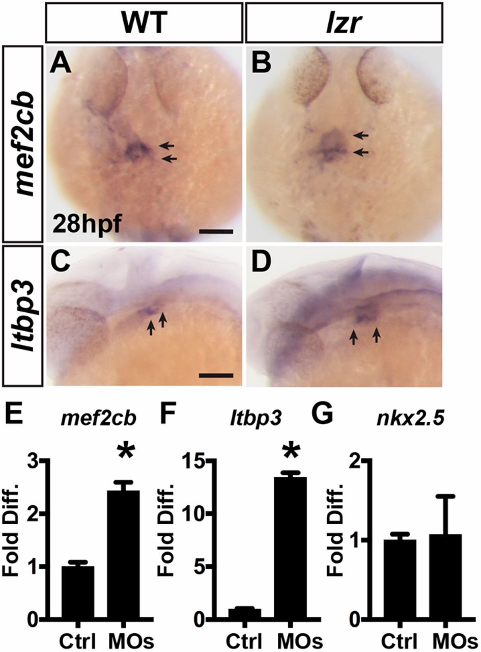
Fig. 5 SHFPs adjacent to the arterial pole of the ventricle are expanded in lzr mutants. (A,B) ISH for mef2cb at 28 hpf. WT (n=7) and lzr (n=6). (C,D) ISH for ltbp3 at 28 hpf. WT (n=7), lzr (n=4). Views are dorsal with anterior up in A and B and lateral with anterior left in C and D. Arrows indicate expression at the arteriole pole of the ventricle. Scale bars: 100 µm. (E-G) RT-qPCR for meb2cb, ltbp3 and nkx2.5 from sorted nkx2.5:ZsYellow+ cells isolated at 28 hpf. Error bars indicate s.e.m. *P<0.05.
Figure Data
Acknowledgments
This image is the copyrighted work of the attributed author or publisher, and
ZFIN has permission only to display this image to its users.
Additional permissions should be obtained from the applicable author or publisher of the image.
Full text @ Development