
|
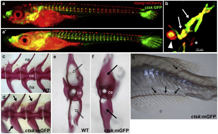
Fig. 1 Osteoclast analysis in Rankl transgenic medaka embryos. (a, a’) Live imaging of macrophages (mpeg:mCherry) and osteoclasts (ctsk:GFP) in rankl:HSE:CFP transgenic medaka embryos at 1 (a) and 3 (a’) days after Rankl induction by heat-shock at 9 days post fertilization. Upon Rankl induction, mpeg:mCherry positive macrophages accumulate at the vertebral column and differentiate into ctsk:GFP expressing osteoclasts. Note that GFP positive cells in the head and caudal fin are cathepsin K expressing cells of unknown identity. (b) High magnification view of vertebral neural arch region showing two macrophages that have differentiated into osteoclasts (ctsk:GFP and mpeg:mCherry double-positive; arrows), and a macrophage that has engulfed a dying osteoclast (arrowhead). Asterisk indicates a non-differentiated macrophage. (c, d) Osteopetrosis phenotype in an osteoclast-deficient, ctsk-mEGFP transgenic medaka, expressing a membrane-localized version of GFP in osteoclasts. Lateral views of Alizarin red stained mineralized matrix in the vertebral column of a wild type (c) and ctsk:mEGFP transgenic medaka (d) at 3 months post fertilization. (e, f) Transverse view of individual vertebra of wild type (e) and ctsk:mEGFP transgenic medaka (f). Regions of over-ossification are indicated with arrows. (g) Trunk of 3 week old ctsk:mEGFP fish; dislocated blood vessels are indicated by arrows. Images (A-B) are courtesy of Phan Quang Tien (Phan and Winkler, unpublished), images (C-G) are taken from To et al. (2015).
Reprinted from Developmental Biology, 457(2), Lleras-Forero, L., Winkler, C., Schulte-Merker, S., Zebrafish and medaka as models for biomedical research of bone diseases, 191-205, Copyright (2019) with permission from Elsevier. Full text @ Dev. Biol.