
|
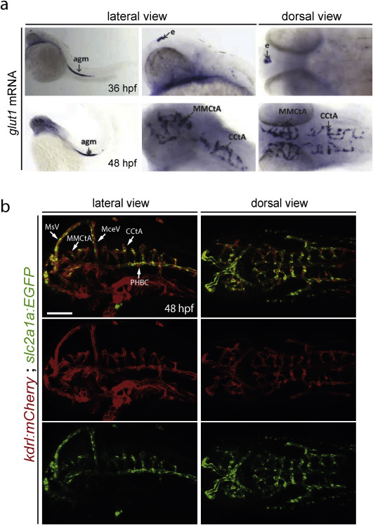
Fig. 3 glut1 mRNA expression labels the brain capillaries of the BBB. a) glut1b mRNA expression in zebrafish embryos. At 36 hpf (upper panels) glut1 expression is detected in the epiphysis (e) and in the aorta-gonad-mesonephros (agm). At 48 hpf (lower panels) glut1 expression becomes prominent in the brain capillaries (MCtAs and CtAs). In situ probe synthesis and hybridization was done according to (Thisse and Thisse, 2008) using the following primers: fwd: GCAGGAGGAACTCAATGCTC, rev+T7; AACGTAATACGACTCACTATAGGGAAACTGGAAGCACATTCCCACT. b) Tg(slc2a1a:EGFP)mu213 reporter line expression in 48 hpf embryos. At 48 hpf glut1b (slc2a1a) is strongly expressed in brain capillaries (MCtAs and CtAs) of Tg(kdrl:ras-mCherry)s896 and in the extracerebral vessels PHBC, BA, BCA, MCeV, MtA, MsVs, but not in the rest of the vessels in the head (lateral view). 8.7 kb of the slc2a1a genomic region upstream of the ATG were cloned upstream of EGFP via Gateway into a Tol2 destination vector (Villefranc et al., 2007) and used for transgenesis as describe previously (Helker et al., 2015). PHBC, primordial hindbrain channel; BA, basilar artery; BCA, basal communicating artery; MCtAs, mesencephalic central arteries; CtAs, central arteries; MCeV, middle cerebral vein; MtA, metencephalic artery; MsV, mesencephalic vein; Scale bar: 100 μm.
Reprinted from Developmental Biology, 457(2), Quiñonez-Silvero, C., Hübner, K., Herzog, W., Development of the brain vasculature and the blood-brain barrier in zebrafish, 181-190, Copyright (2019) with permission from Elsevier. Full text @ Dev. Biol.