IMAGE

Figure 8

ID
ZDB-IMAGE-200413-24
Source
Figures for Sie et al., 2020
Image
Figure Caption

Figure 8

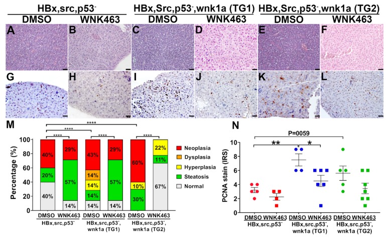
Effect of WNK1 pathway inhibitors on HCC in transgenic fish overexpressing endothelial cell-specific wnk1a. (AF) Representative images of H&E-stained liver sections from HBx, src(p53-) fish and two lines (TG1 and TG2) of HBx, src(p53-) transgenic fish overexpressing wnk1a in endothelia. Scale bar = 20 μm. (G–L) Representative images of PCNA staining of liver sections from fish as above. (M) Distribution of histopathological diagnosis of H&E-sections as shown in panels (A–F). The statistical significance was analyzed by Chi-square test. ****: p ≤0.0001. (N) Average immunoreactive score (IRS) of proliferating cell nuclear antigen (PCNA) staining as shown in panels (G–L). The statistical significance was analyzed by unpaired Student’s t-test. *: 0.01< p ≤0.05; **: 0.001< p ≤0.01.

Acknowledgments
This image is the copyrighted work of the attributed author or publisher, and ZFIN has permission only to display this image to its users. Additional permissions should be obtained from the applicable author or publisher of the image. Full text @ Cancers