IMAGE

Fig 1

ID
ZDB-IMAGE-200406-50
Source
Figures for Wang et al., 2020
Image
Figure Caption

Fig 1 Construction of in vivo circadian reporter in zebrafish.

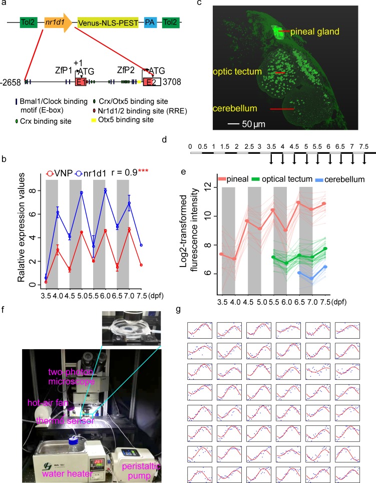
(a) The upper graph shows the schematic of nr1d1:VNP construct design. The lower graph shows a magnified view of the nr1d1 promoter sequence used for driving the circadian expression of VNP. The putative RRE (Nr1d1/2 binding site), E-box (Bmal1/Clock binding site), Crx, Otx5, and Crx/Otx5 binding sites were indicated by red oval, blue rectangle, light green oval, yellow rectangle, and dark green oval, respectively. (b) Plot of real-time PCR results of nr1d1 and Venus-pest expression. Each time point has 2 replicates, and each replicate is the pool of 7–10 fish. The dots show the original value, while the solid lines and the bars represent mean ± SEM. (c) Whole-brain two-photon image shows the spatial distribution of nr1d1:VNP-positive cells in different brain regions at 7.5 dpf. (d) Experimental design to examine the developmental dynamics of nr1d1:VNP expression in the whole zebrafish brain. (e) Single-cell tracing result of nr1d1:VNP-positive cells in the pineal gland (red), the optic tectum (green), and the cerebellum (blue) for one fish. The cells were tracked from 3.5 dpf in the pineal gland, 5.5 dpf in optic tectum, and 6.5 dpf in cerebellum, respectively. Each thin line represents one cell, while the thick line represents the mean value of cells in each brain region. (f) Illustration of the setup for time-lapse imaging. The fish were fixed in a chamber with circulating egg-water without anesthesia at temperature of 28 ± 0.5°C. (g) Single-cell tracing results of all 48 nr1d1:VNP-positive cells imaged at 1-hour resolution. The blue dots represent the original fluorescence signals, while the solid red line represents the smoothed curve fitted by the cosine functions. The numerical values for panels b, e, and g are in S1 Data. dpf, days postfertilization; PA, poly(A) site; Tol2, transposable element of Oryzias latipes 2; VNP, Venus-NLS-PEST.

Acknowledgments
This image is the copyrighted work of the attributed author or publisher, and ZFIN has permission only to display this image to its users. Additional permissions should be obtained from the applicable author or publisher of the image. Full text @ PLoS Biol.