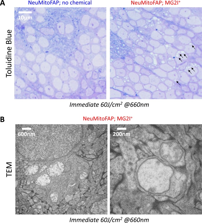
IMAGE

Figure 5-S1

ID
ZDB-IMAGE-200406-263
Source
Figures for Xie et al., 2020
Image
Figure Caption

Figure 5-S1 Widespread disruption of mitochondrial morphology in NeuMitoFAP zebrafish exposed to MG2I and far-red light.

NeuMitoFAP zebrafish larvae were treated with MG2I or no chemical from 3 to 5 dpf and then exposed to 60 J/cm2 light at λpeak=661 nm, following which they were fixed immediately for electron microscopy. (A) Toluidine Blue-labeled thin sections of the telencephalon, showing the widespread appearance of lucent areas in the cytoplasm of NeuMitoFAP-MG2I neurons following light exposure (right panel; examples of lucent areas are indicated with arrows). These abnormalities were not present in controls that were not treated with MG2I (left panel). (B) Transmission electron microscopy images showing that the lucent areas correspond to severely damaged mitochondria that were swollen and lacked cristae. Multiple abnormal mitochondria were frequently seen within the same cell (left). Severely damaged mitochondria were also apparent within axons and terminals (right) corresponding to the fragmented and swollen axonal mitochondria seen in Figure 5A.

Acknowledgments
This image is the copyrighted work of the attributed author or publisher, and ZFIN has permission only to display this image to its users. Additional permissions should be obtained from the applicable author or publisher of the image. Full text @ Elife