
|
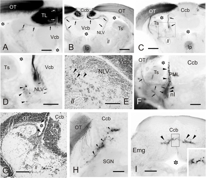
FIGURE 7
Labeled cells and fibers after direct DiI application to TL. Photomicrographs of transverse sections caudal to the tracer application point showing labeled structures observed after homolateral DiI application to intermediate TL. Ipsilateral side is to the left.