IMAGE

FIGURE 3

ID
ZDB-IMAGE-200406-110
Source
Figures for Folgueira et al., 2020
Image
Figure Caption

FIGURE 3

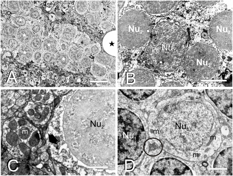
Fine cell structure of the adult TL. (A–D) Electron micrographs showing the main three nucleus types found in TL. (A) Low magnification electron micrograph showing a cell cluster with medium-sized round nuclei with partially condensed chromatin (Nu1, white star) and surrounded by a dense neuropil. (B) Detail of a medium-sized nucleus (Nu1) surrounded by smaller nuclei (Nu2 and Nu2). (C) Detail of a small-sized cell nucleus with non-homogeneously condensed chromatin (Nu2) showing the exit of the axon (black arrow) from the cell body. (D) Nucleus (Nu3) with pale chromatin that belongs to the large-sized cells among other nuclei with more condensed chromatin (Nu1). Note also the small compact bundle of non-myelinated axons (circle). m, mitochondria; black stars, blood vessels. Scale bars: 5 μm (A), 2 μm (B,D), 1 μm (C).

Acknowledgments
This image is the copyrighted work of the attributed author or publisher, and ZFIN has permission only to display this image to its users. Additional permissions should be obtained from the applicable author or publisher of the image. Full text @ Front. Neural Circuits