
|
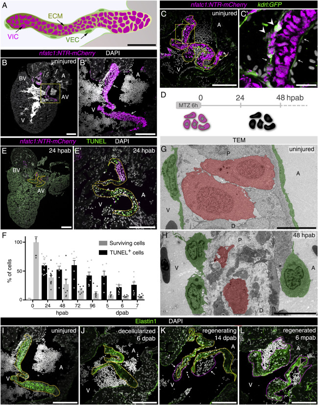
Fig. 1 Decellularization of the Zebrafish Atrio-Ventricular Valve Triggers a Regenerative Program Leading to a New Valve (A) Schematics of the zebrafish AV valve leaflet depicting the lining VECs and the ECM-embedded VICs. (B and B′) Cryosection of Tg(nfatc1:NTR-mCherry) heart showing NTR-mCherry expression in VICs in the AV and bulbo-ventricular (BV) valves (arrows point to nfatc1− VECs). Boxed area shown in (B′). (C and C′) A minority of VECs are NTR-mCherry+ (arrowheads), while most kdrl:GFP+ cells are NTR-mCherry− (arrows; n = 4). Boxed area shown in (C′). (D) Ablation protocol using the NTR-Mtz system (D). (E and F) TUNEL detection (E and E′) and quantification (F) in ablated hearts show apoptotic cells only in the valve leaflets. Plot values represent means ± SEM. (G and H) TEM images of uninjured (G; n = 3) and 48 hpab (H; n = 3) valve sections showing the morphology of the lining VECs (green) and VICs (red) before and after ablation. (I–L) Representative images of the AV valve regeneration process illustrating the uninjured valve (I; same section as in Figures 1B and 1B′; n = 8), tissue decellularization (J; n = 16), formation of new valve leaflets (K; arrows point to new valve cells; n = 8), and completion of regeneration (L; n = 10) with ECM labeled by Elastin1 antibody. Yellow and pink dashed lines delineate the old and new valve leaflets, respectively. A, atrium; V, ventricle; P, proximal; D, distal. Scale bars: (A) 50 μm; (B and E) 200 μm; (B′), (C), (E′), and (I–L) 100 μm, (C′) 20 μm; and (G and H) 10 μm. See also Figure S1; Videos S1, S2, S3, and S4.
Reprinted from Developmental Cell, 52(1), Bensimon-Brito, A., Ramkumar, S., Boezio, G.L.M., Guenther, S., Kuenne, C., Helker, C.S.M., Sánchez-Iranzo, H., Iloska, D., Piesker, J., Pullamsetti, S., Mercader, N., Beis, D., Stainier, D.Y.R., TGF-β Signaling Promotes Tissue Formation during Cardiac Valve Regeneration in Adult Zebrafish, 9-20.e7, Copyright (2019) with permission from Elsevier. Full text @ Dev. Cell