
|
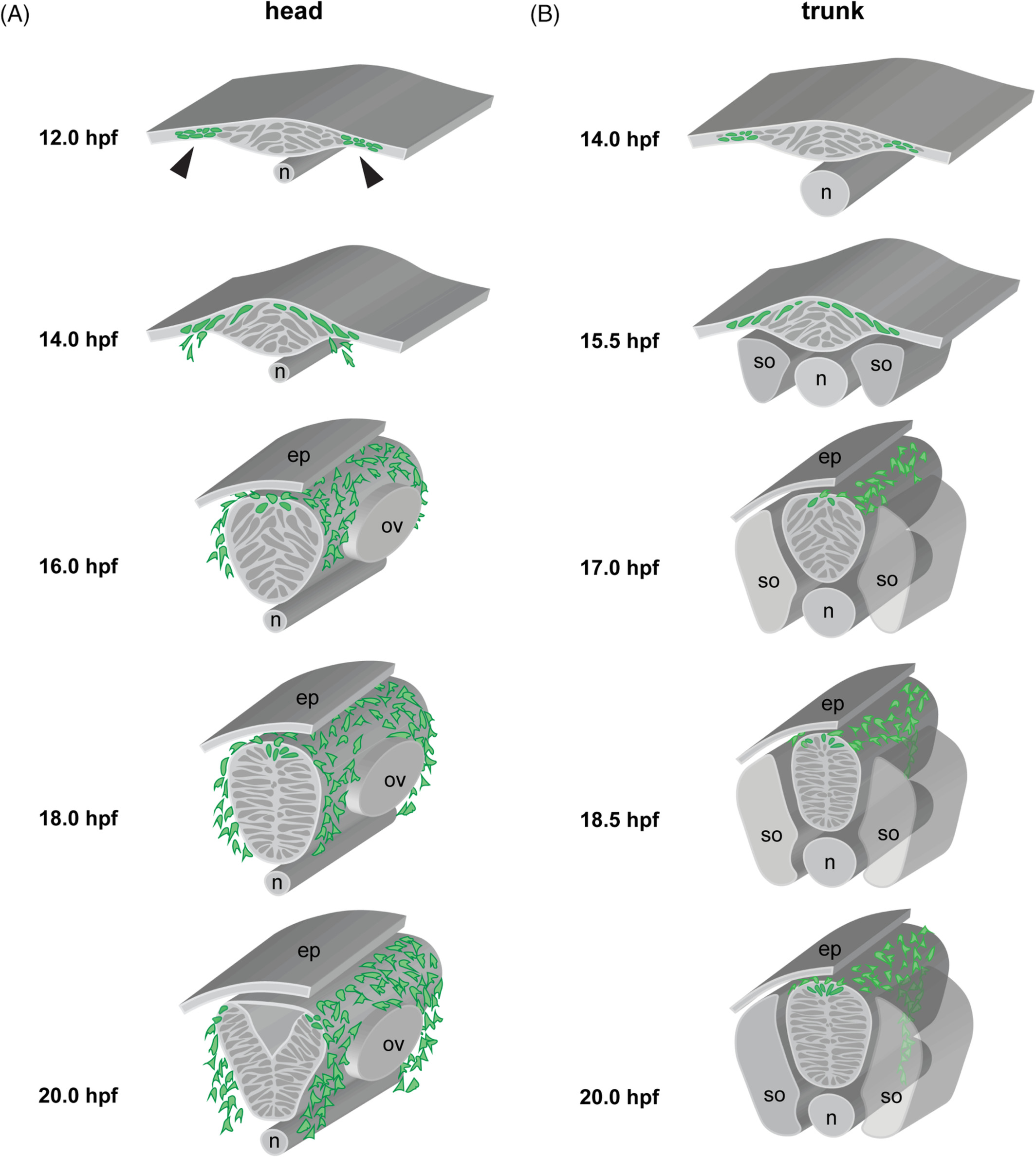
Fig. 2 Zebrafish neurulation and neural crest migration pathways. A, Cranial neurulation and neural crest migration, schematized at the rhombomere (r)4‐6 level, showing neural crest cells (green) migrating anterior and posterior of the otic vesicle towards pharyngeal arch (PA)2 and PA3, respectively. Stages are indicated, with laterally segregated cells apparent at 12 hpf (arrowheads), convergence movements forming the neural keel by 14 hpf, a neural rod at 18 hpf (with a clear midline now established), and finally a neural tube at 20 hpf. Note that the anterior terminus of the notochord lies under r4 in the zebrafish. B, Trunk neurulation and neural crest migration, schematized at the somite 7/8 level, showing neural crest cells (green) migrating on the medial pathway adjacent to the center of each somite. Stages are indicated. n = notochord; ep = epidermis; ov = otic vesicle; s = somite