IMAGE

Fig 5

ID
ZDB-IMAGE-200325-62
Genes
Antibodies
Source
Figures for Takemoto et al., 2020
Image
Figure Caption

Fig 5 Homologous pairing is impaired in <italic>sycp2</italic> mutant spermatocytes.

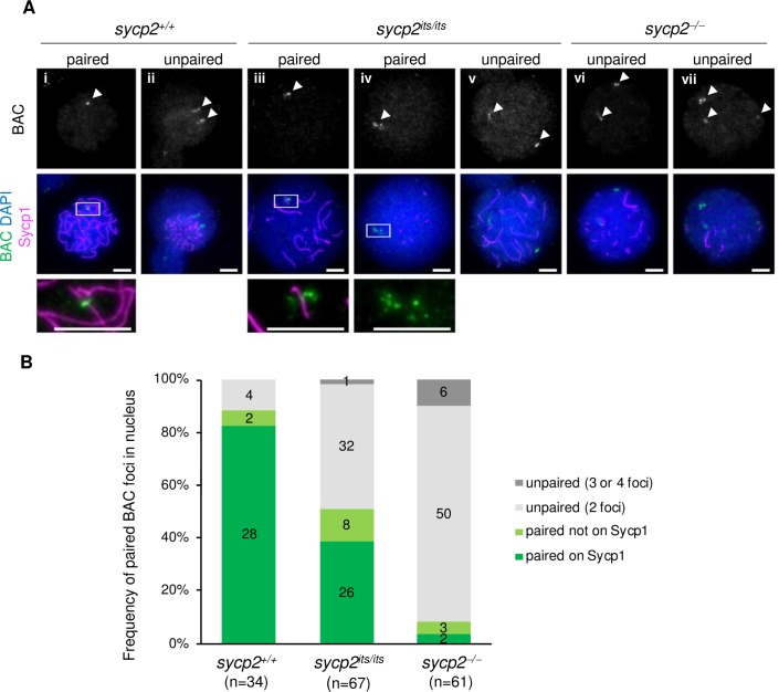
A: Fluorescent in situ hybridization with a BAC probe in sycp2+/+, sycp2its/its and sycp2-/- spermatocytes. The regions outlined with white in A-i, A-iii and A-iv are shown at a higher magnification at the bottom. White arrowheads indicate BAC-probe-stained foci. A-i, A-iii and A-iv show nuclei with paired BAC foci with (A-i and A-iii) and without (A-iv) colocalization on an Sycp1 fragment. A-ii, A-v, A-vi and A-vii show nuclei with multiple unpaired BAC foci. Scale bars, 5 μm. B: Quantification of pairing of BAC foci in sycp2+/+ (n = 34), sycp2its/its (n = 67) and sycp2-/- (n = 61) spermatocytes. The percentage of nuclei stained for one to four BAC foci is shown for each genotype. When BAC signals in a nucleus were observed as one focus or two foci with an intervening distance of <3 μm, they were considered to be paired, a localization on Sycp1 filaments was also evaluated. Proportions corresponding to one BAC focus with localization (paired on Sycp1) and without localization (paired not on Sycp1) on Sycp1 filaments are shown in dark and light green, respectively. Chromosomal spreads of one (wild-type) or two (sycp2its/its and sycp2-/-) individual fish were used for counting.

Figure Data
Acknowledgments
This image is the copyrighted work of the attributed author or publisher, and ZFIN has permission only to display this image to its users. Additional permissions should be obtained from the applicable author or publisher of the image. Full text @ PLoS Genet.