IMAGE

Fig. 4

ID
ZDB-IMAGE-200325-54
Source
Figures for Ren et al., 2020
Image
Figure Caption

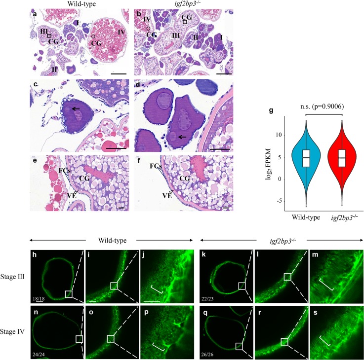
Fig. 4 Oogenesis is largely unaffected in <italic>igf2bp3</italic> homozygous mutant (<italic>igf2bp3</italic><sup>−/−</sup>) zebrafish.

a, b H&E-stained ovary sections indicated cortical granule movement toward the cortex, structure of the vitelline envelope, and somatic follicle cells surrounding stage I–IV oocytes of wild type and igf2bp3−/− zebrafish. CGs cortical granules. Square box indicates CGs located centrally in young oocytes and round box indicates CGs in the cortex at late stage; Scale bars:100 μm. c, d The normal polarization of stage Ib oocytes as indicated by the presence of the Balbiani body (black arrowheads) in igf2bp3−/− zebrafish compared with the wild type. Scale bars: 50 μm. e, f H&E-stained stage III oocytes revealed cortical granule movement toward the cortex in wild type and igf2bp3−/− zebrafish. VE vitelline envelope (triangle box); FCs follicle cells (two straight lines). Scale bars: 20 μm. g Violin and box plots depicting the expression levels of maternal genes in unfertilized eggs. Lower/middle/upper position in box plots indicated 25/50/75% quantile, respectively. The violin width shows the gene density. Two-sided Wilcoxon and Mann–Whitney test was used. n.s. no significant. hs F-actin expression in wild type and igf2bp3 mutant oocytes. The inset showed a zoom-in of the boxed region. i, l, o, r In the subcortical regions of stage III–IV oocytes, the actin filaments became clearly distributed in both wild type and igf2bp3 mutant oocytes. j, m, p, s High magnification images of actin filaments in the oocyte cytoplasm. The actin columns were arranged from the inner to the outer across the cortical cytoplasm and thereby the column was arranged parallel to each other (brackets). Scale bars: 100 μm in h, k, n, q; 25 μm in i, l, o, r; 10 μm in j, m, p, s.

Figure Data
Acknowledgments
This image is the copyrighted work of the attributed author or publisher, and ZFIN has permission only to display this image to its users. Additional permissions should be obtained from the applicable author or publisher of the image. Full text @ Commun Biol