
Image
|
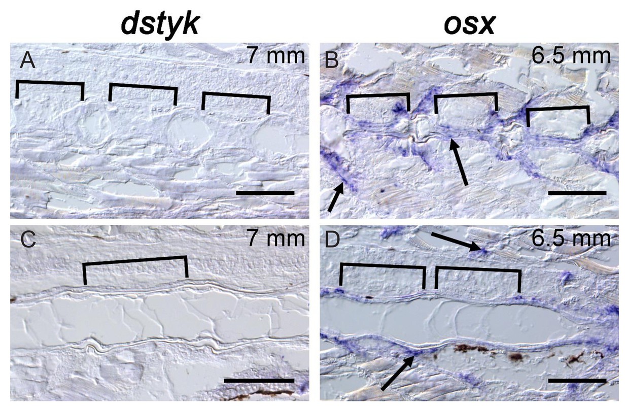
Figure Caption
Fig. 10-S1
dstyk expression is not detectable at late stages in the notochord or in osteoblasts surrounding the notochord.
( A,C) In situ hybridization for dstyk at 7 mm stage. Sagittal sections were taken at the periphery of the notochord ( A) and across the center of the notochord ( C). Brackets indicate the mineralized centra. ( B, D) In situ hybridization for osx at 6.5 mm standard length. Sagittal sections were taken at the periphery of the notochord ( B) and across the center of the notochord ( D). Brackets indicate the mineralized centra. Brackets indicate mineralized centra. Arrows point to osteoblasts. Scale bar = 100 µm.
Acknowledgments
This image is the copyrighted work of the attributed author or publisher, and
ZFIN has permission only to display this image to its users.
Additional permissions should be obtained from the applicable author or publisher of the image.
Full text @ Elife