
IMAGE
Fig. 7-S1
- ID
- ZDB-IMAGE-200325-187
- Publication
- Bagwell et al., 2020 - Notochord vacuoles absorb compressive bone growth during zebrafish spine formation
- All Figures
- Figures for Bagwell et al., 2020
Image
|
Figure Caption
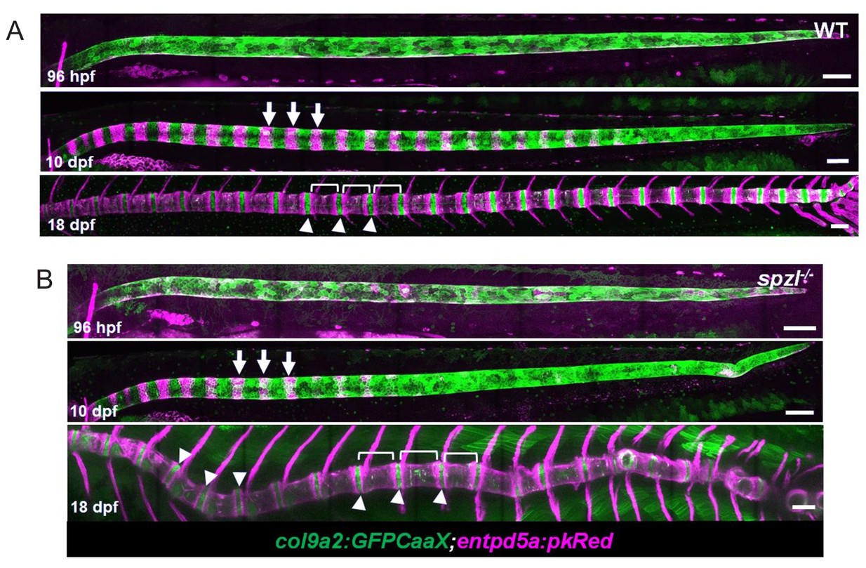
Fig. 7-S1
Notochord segmentation occurs normally in spzl mutants.
( A–B) Maximum intensity projections of live confocal images from WT and spzl-/- larvae during notochord segmentation. ( B) Arrows point to mineralizing segments labeled with entpd5a:PkRed. Brackets highlight vertebrae. Arrowheads mark the intervertebral domains which are labeled with col9a2:GFPCaaX. Scale bar = 100 µm.
Acknowledgments
This image is the copyrighted work of the attributed author or publisher, and
ZFIN has permission only to display this image to its users.
Additional permissions should be obtained from the applicable author or publisher of the image.
Full text @ Elife