
|
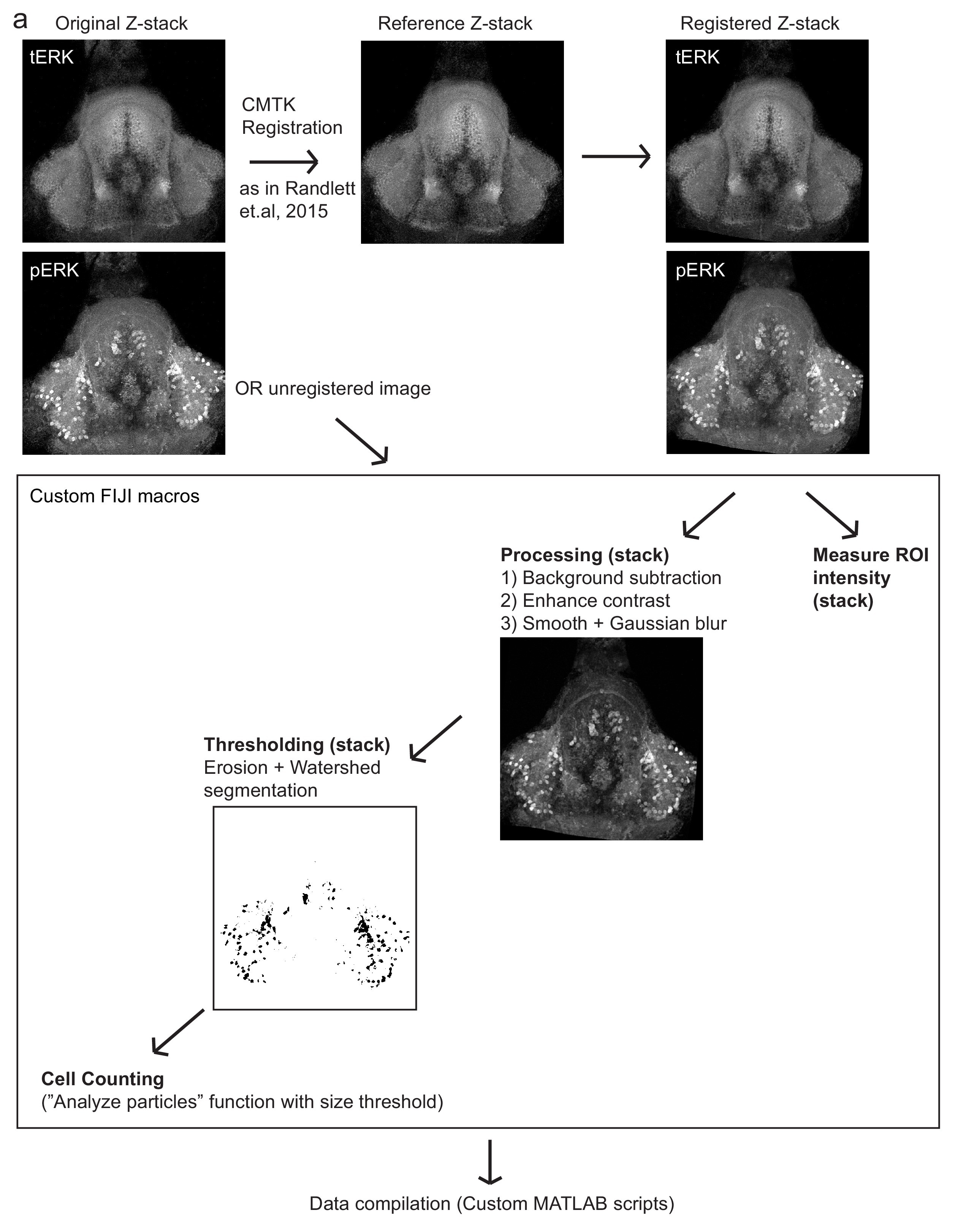
Fig. 1-S4
Automated quantification of pERK-positive (active) cells.
( a) Method by which pERK-positive (‘active’) cell count were determined in a high-throughput manner. Brain z-stacks obtained from confocal microscopy are registered with a selected reference brain within the same dataset, using the tERK channel, though in experiments where tERK staining was not performed, unregistered images were used (for which individual ROIs have to be defined for each image). A series of processing steps were uniformly applied to segment pERK-positive cells, which were selected using a manually optimized threshold across the entire dataset. Cell counts were obtained using the Analyze Particles algorithm within the Fiji software.