
|
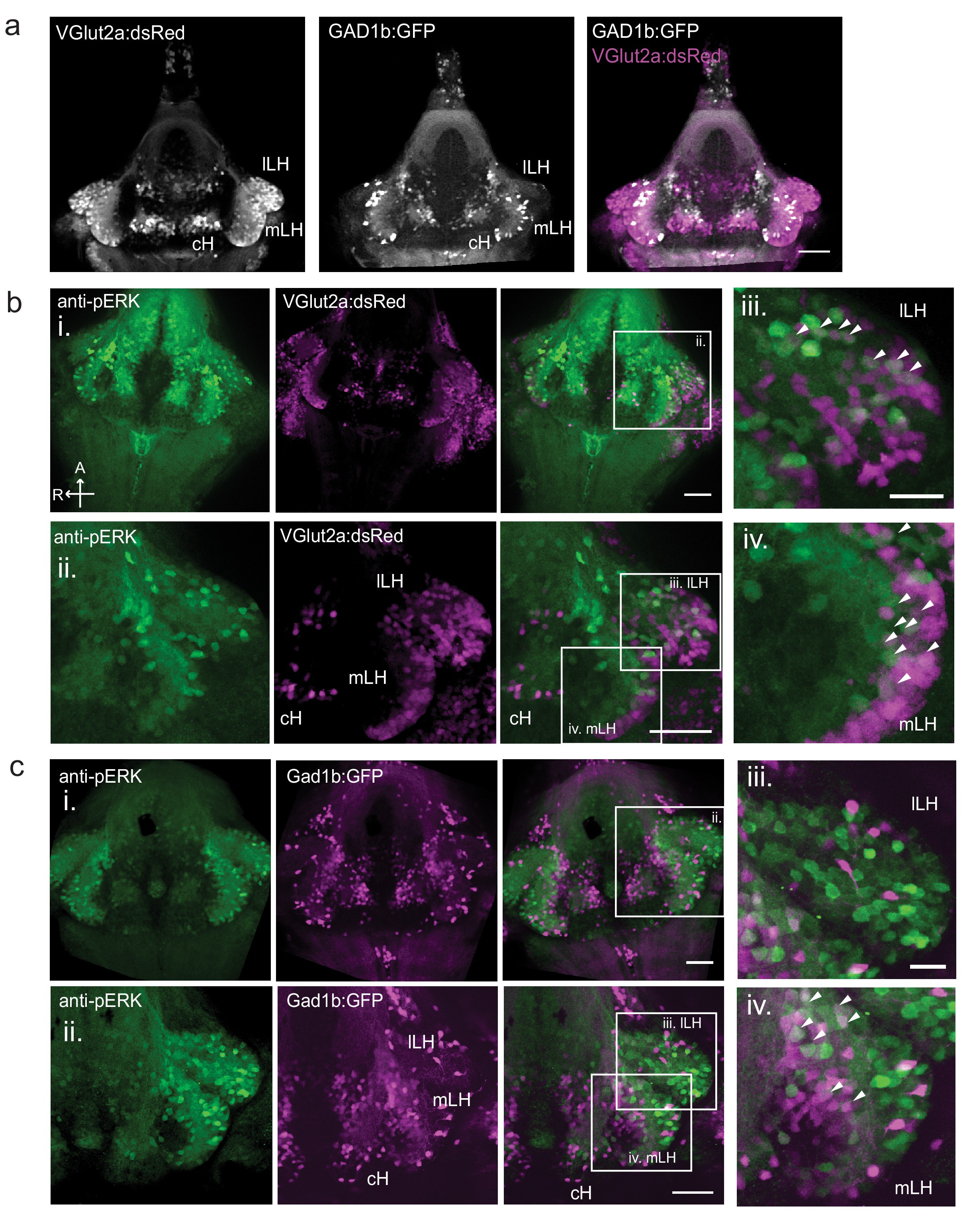
Fig. 1-S2
Characterization of neuronal transmitter types in the zebrafish lateral hypothalamus.
( a) Glutamatergic and GABAergic neuron distribution in the hypothalamus. Tg(VGlut2a:dsRed) and Tg(GAD1b:GFP) transgenic fish were dissected, imaged and registered onto a common reference hypothalamus. All fish in this figure were food-deprived for 2 hr and fixed for analysis after 15 min of feeding. ( b) Glutamatergic cells, labeled by Tg(VGlut2a:dsRed), overlap with active (pERK-positive) neurons in both the lLH and outer rim of the mLH. (i) Z-projection of hypothalamus. (ii) Higher magnification images of LH (iii-iv) Inset showing overlap of lLH and outer rim of mLH with glutamatergic cells. ( c) GABAergic cells, labeled by Tg(Gad1b:GFP), overlap with active neurons in the inner rim of the mLH but not the lLH. (i) Z-projection of hypothalamus. (ii) Higher magnification images of LH showing a subset of z-planes. (iii-iv) Inset showing overlap of inner rim of mLH with GABAergic cells. White arrows point to examples of overlapping cells. All fish were mounted ventral side up. Scale bar (i and ii) = 50 μm. Inset (iii and iv) scale bar = 20 μm.