
Fig. 4
- ID
- ZDB-IMAGE-200323-22
- Publication
- Caceres et al., 2019 - Frizzled 4 regulates ventral blood vessel remodeling in the zebrafish retina
- All Figures
- Figures for Caceres et al., 2019
|
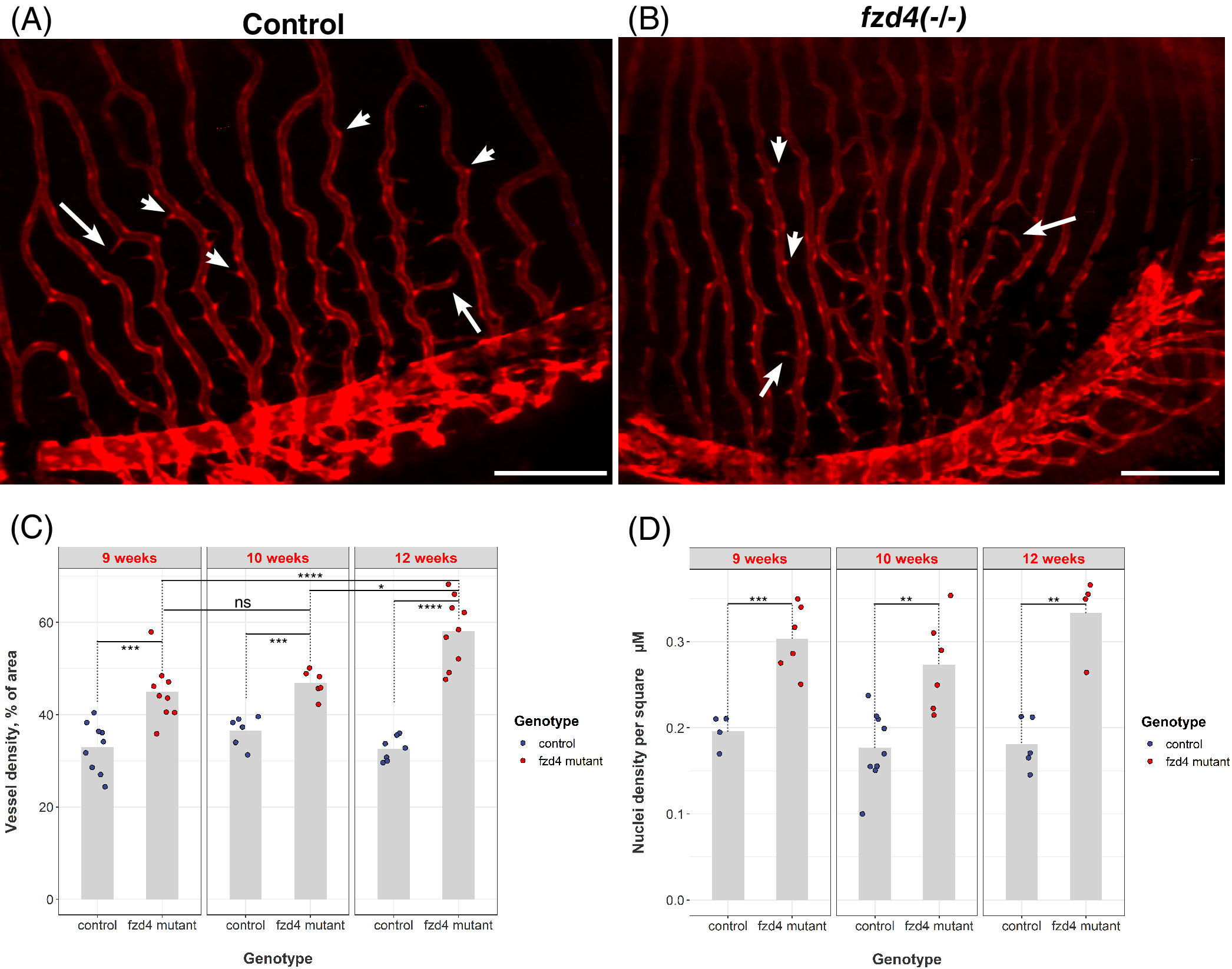
Fig. 4 Loss of fzd4 results in increased retinal vessel density and endothelial cell proliferation. A‐B, Thin cellular projections (arrows) emanating from the red foci of the endothelial cell bodies (arrowheads) to form a kinked and branching pattern in the vasculature are detected in both, A, control and B, fzd4 mutant retinas. Green fluorescent protein (GFP) (red) marks retinal vasculature. Scale = 200 μm. C‐D, Quantification of cellular fusion and proliferation shows a progressive increase over time in vessel density of fzd4 mutants compared to controls and itself at earlier time points (compare fzd4 week 9 to fzd4 week 12). Furthermore, this increase in vessels density results from increase in endothelial proliferation as shown in D