
Fig. 1
- ID
- ZDB-IMAGE-200323-19
- Publication
- Caceres et al., 2019 - Frizzled 4 regulates ventral blood vessel remodeling in the zebrafish retina
- All Figures
- Figures for Caceres et al., 2019
|
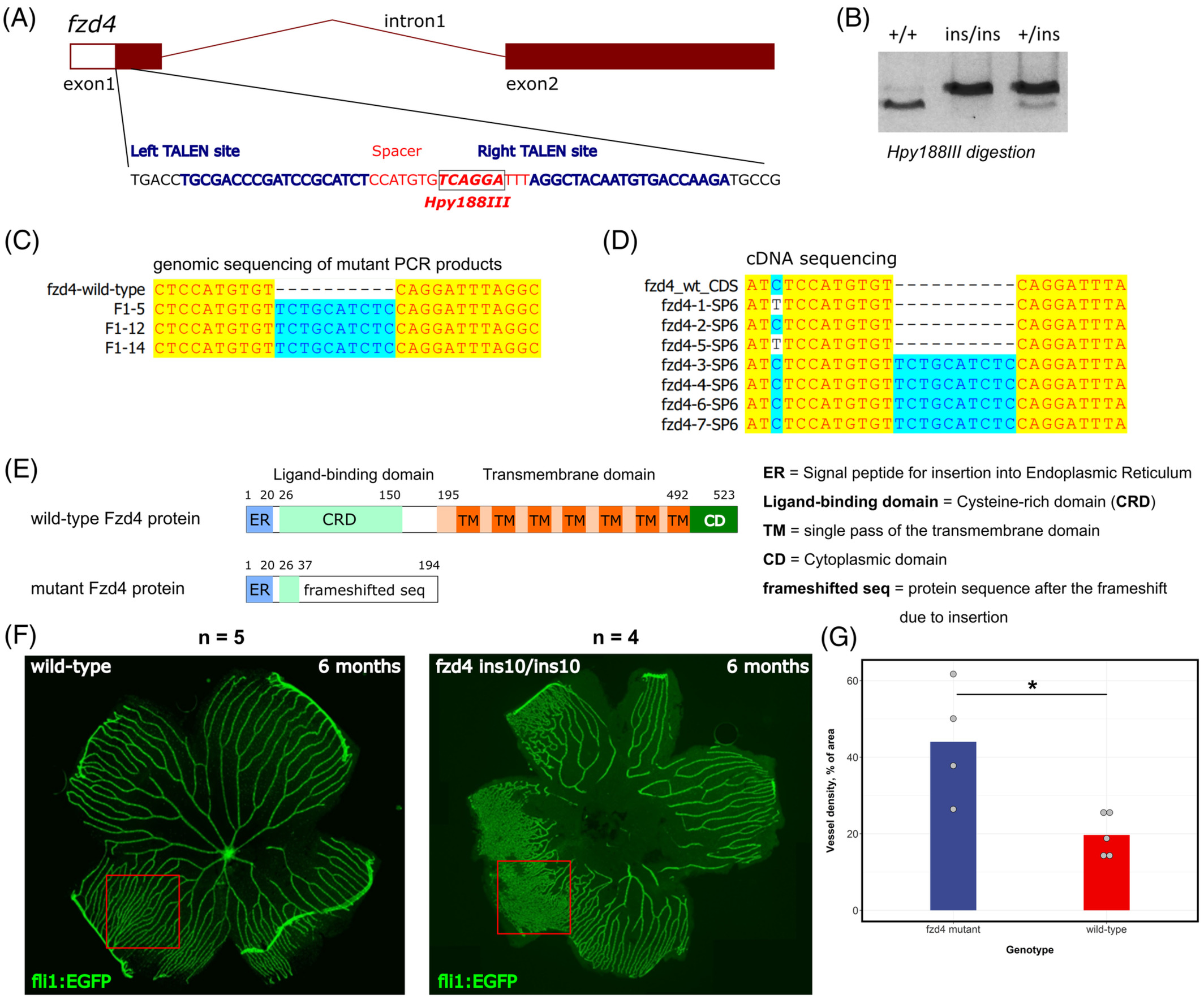
Fig. 1 Generation of the fzd4 ins10 mutant and initial characterization of its retinal vasculature phenotype. A, The exon‐intron structure of the zebrafish fzd4 gene is shown with a zoomed‐in section of exon 1, which was targeted by TALENs. The targeted part of exon 1 contains left and right binding sites for TALEN proteins and the spacer sequence between them is the mutagenesis target. The site of the Hpy188III enzyme within the spacer is indicated by a label, box, and bold font. B, An example of the 10‐nucleotide insertion mutant (ins10) genotyping by first amplifying PCR products of the target site from wild‐type (+/+), heterozygous (+/ins) and homozygous mutant (ins/ins) animals followed by Hpy188III digestion and agarose gel electrophoresis. C, Sequencing results of the Hpy188III‐undigestable PCR products derived from F1 mutation‐carrier zebrafish show clear evidence of the 10‐nucleotide insertion at the TALEN‐targeted site. D, Sequencing of individual plasmid clones containing cDNA fragments from the RNA of fzd4 +/ins10 incrossed embryos. Among eight clones analyzed, one was empty (not shown), four had the insertion, and three had the wild‐type version of the cDNA. E. Domain diagrams and sizes are shown for the wild‐type (523 amino acids) and ins10 mutant (194 amino acids in total, with 37 N‐terminal amino acids of Fzd4). Fzd4 proteins predicted from cDNA sequencing. The following domains and structures are shown: ER = Signal peptide for insertion into endoplasmic reticulum, CRD = cysteine‐rich domain (the Norrin ligand‐binding domain), TM = single pass of the transmembrane domain, CD = cytoplasmic domain, frameshifted seq = protein sequence after the frameshift due to insertion. F, Imaging of dissected retinal vasculature of 6‐month old wild‐type control (Tg[fli1:EGFP]AB) and fzd4 ins10/ins10 mutant fli1:EGFP transgenic zebrafish. Note the disorganized vasculature in the mutant. G. Measurement of vessel density shows an increase in the vascular network of fzd4 mutant fish compared to its control counterpart