
|
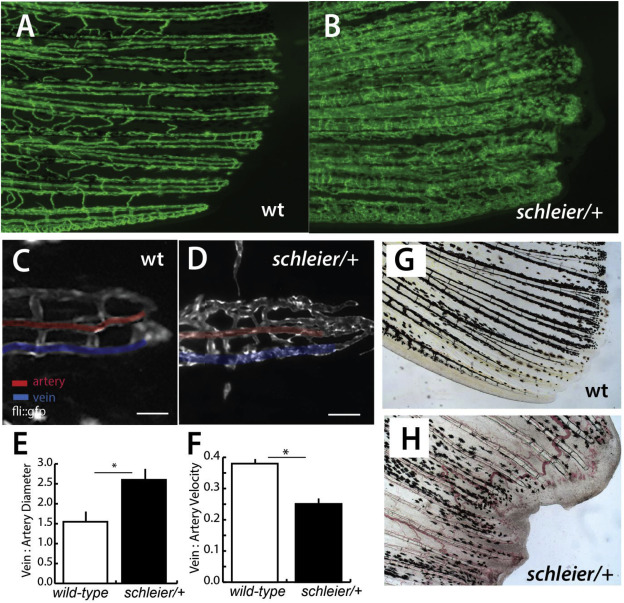
Fig. 2 Schleier mutation highlights role of vascular tone associated with altered proportionality in zebrafish fins. A, B) Pattern of vascularization in caudal fins of adult wild-type (wt) and schleier heterozygous fish visualized by Tg(fli1:egfp) transgenic expression of endothelial cells. C, D) Close up of caudal fin tip showing venous and arterial pattern variation in the schleier mutant associated with increased number of endothelial cells. Veins and arteries pseudo-colored blue and red, respectively; scale bar equals 100 μM. E) Measure of vascular dilation in schleier caudal fin tips. Mean ± standard error of mean (SEM) is shown. *, p < 0.05 by ANOVA. N = 3 fish per genotype; mean of 5 artery and 5 vein diameter measurements per fish. F) Quantitation of vascular flow in fin tip in arteries compared with veins. Mean ± standard error of mean (SEM) is shown *, p < 0.05 by ANOVA. N = 3 fish per genotype; mean of 10 arterial red blood cells and 10 venous red blood cells per fish. G, H) Excessive vascularization and blood pooling in the schleier mutant fin tip (H) compared with wild-type fin tip (G).
Reprinted from Developmental Biology, 456(2), Lanni, J.S., Peal, D., Ekstrom, L., Chen, H., Stanclift, C., Bowen, M.E., Mercado, A., Gamba, G., Kahle, K.T., Harris, M.P., Integrated K+ channel and K+Cl- cotransporter function are required for the coordination of size and proportion during development, 164-178, Copyright (2019) with permission from Elsevier. Full text @ Dev. Biol.