
|
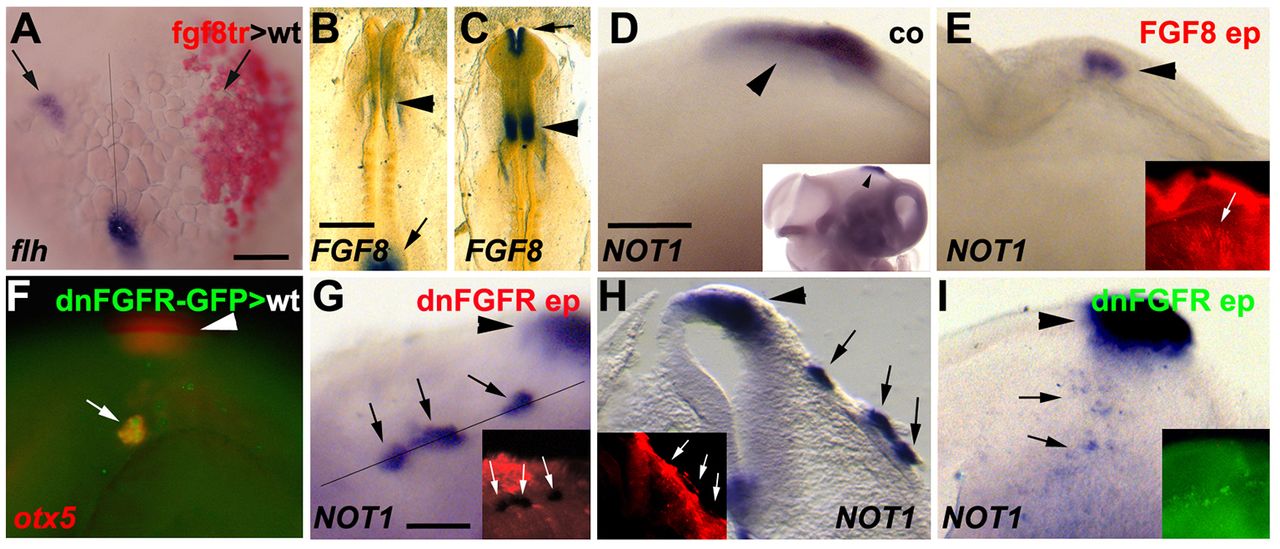
Fig. 7 FGF signalling antagonises pinealogenesis. (A) Bud stage zebrafish embryo (anterior points to the top) containing transplanted fgf8-overexpressing cells in the right side of the neural plate (red), labelled using in situ hybridisation for flh (blue). flh-positive pineal progenitors are present on the right side (arrows; n=7/7). (B,C) Chick embryos (anterior towards the top) at HH8.5 (B) and HH9.5 (C) stained using in situ hybridisation for expression of FGF8 (n=6; two independent experiments). Expression is present in the midbrain-hindbrain boundary in B (arrowhead; the arrow marks expression in the tailbud region). Expression is present in the anterior neural folds (arrow) and midbrain-hindbrain boundary (arrowhead) in C. (D) HH18 chick embryo stained using in situ hybridisation for NOT1 (lateral view of diencephalon, anterior is rightwards). Inset shows same embryo at lower magnification. (E) Chick embryo electroporated at HH10 with FGF8 and eGFP, fixed after 36 h of incubation and stained by in situ hybridisation for expression of NOT1 (blue) and by immunohistochemistry for GFP (red, inset). Arrowheads mark the pineal organ; the arrow in the inset marks the location of electroporated cells at some distance from the pineal organ. NOT1 is downregulated in E compared with D (n=11/15; two independent experiments). (F) Zebrafish embryo (anterior is leftwards, dorsal towards the top) transplanted at 30% epiboly stage with heat shock-inducible dnfgfr-transgenic donor cells (green), heat-shocked at shield stage, fixed at 24 hpf and labelled by in situ hybridisation for otx5 (red). Ectopic pineal progenitors are induced following heat shock (n=9/12). Arrowhead highlights otx5-positive pineal organ; arrow indicates ectopic otx5-positive cells. (G) Lateral view (anterior towards the right) of a chick embryo electroporated at HH8 with dnFGFR and eGFP, and stained after 24 h of incubation by in situ hybridisation for NOT1 (blue) and by immunohistochemistry for GFP (red, inset). Arrowhead indicates NOT1-positive pineal organ; black and white arrows highlight ectopic NOT1-positive cells. (H) Oblique section of embryo along the line indicated in G. Arrowhead indicates pineal organ; arrows indicate clusters of NOT1-expressing cells in superficial ectoderm (n=6/13). (I) Lateral view of chick embryo electroporated at HH8 with dnFGFR and eGFP into the right anterior neural fold, and stained for NOT1 after 36 h of incubation (arrowhead indicates pineal organ). Inset shows GFP fluorescence before in situ hybridisation. There is ectopic induction of NOT1-positive pineal progenitor cells in electroporated neural tube (arrows; n=7/27). Scale bars: 50 µm in A for A-F; in B, 500 µm for B,C; in D, 200 µm for D,E,I; 100 µm in G for G,H.