IMAGE

Figure 1.

ID
ZDB-IMAGE-200316-4
Source
Figures for Hung et al., 2020
Image
Figure Caption

Figure 1.

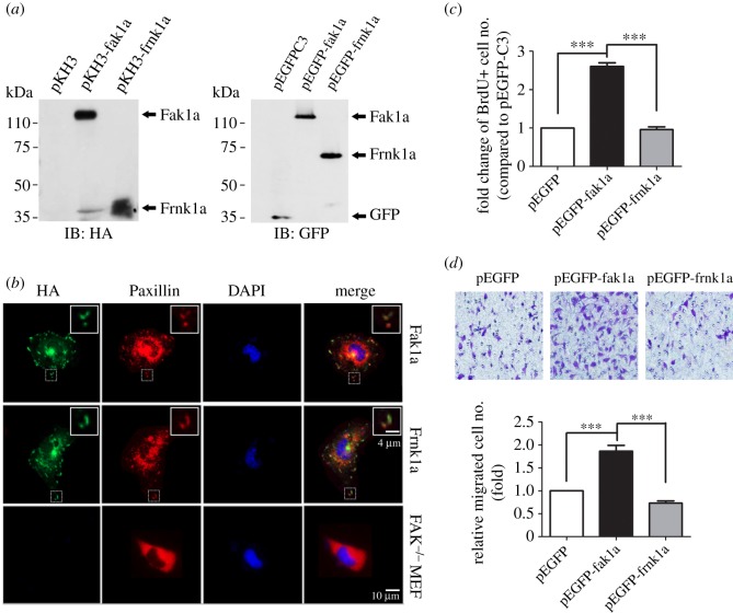
Zebrafish Fak1a is functionally conserved with mammalian FAKs. (a) Zebrafish Fak1a and Frnk1a were successfully expressed in 293T cells as characterized by immunoblotting (IB) using anti-HA (left) and anti-green fluorescent protein (GFP) antibodies (right). (b) Zebrafish Fak1a and Frnk1a with an HA tag were expressed in FAK−/− MEF cells as revealed by HA immunostaining shown in green (left column). They were present in focal contacts (boxed), and an enlarged image is shown at the top right corner of the respective panel. Focal contacts and cell nuclei were visualized by Paxillin (second column) and DAPI staining (third column), respectively. Merged images for Fak, Paxillin and DAPI staining are presented in the right column with enlarged insets for the boxed regions to better show the co-localization of Fak and paxillin in focal contacts. Cells were transfected with pEGFP-C3 vector only, fak1a or frnk1a to examine cell proliferation (c) and cell migration (d) as determined by a BrdU incorporation assay and Bodyen chamber migration assay, respectively (n = 3; ***p < 0.001). Detailed protocols are described in ‘Material and methods'.

Acknowledgments
This image is the copyrighted work of the attributed author or publisher, and ZFIN has permission only to display this image to its users. Additional permissions should be obtained from the applicable author or publisher of the image. Full text @ Open Biol.