IMAGE

Figure 10.

ID
ZDB-IMAGE-200316-1
Source
Figures for Hung et al., 2020
Image
Figure Caption

Figure 10.

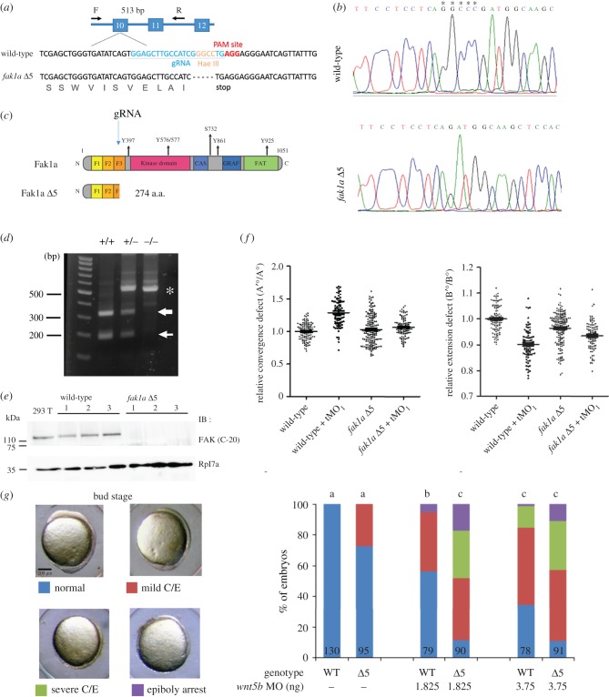
CRISPR/Cas9-mediated deletion of Fak1a results in mild gastrulation defects due to compensatory wnt5b expression. (a) The fak1a gene structure is shown with indicated exons in blue boxes. The target sites of a primer pair (F, forward primer; R, reverse primer) for amplifying a mutation detection 513-bp PCR fragment are indicated by arrows. The sense strand of wild-type fak1a is shown (upper strand). Exon 10 contains a PAM site shown in red. A gRNA target site of the exon 10 PAM site is labelled in blue. The Hae III site is shown in orange. The resulting sense strand of the fak1a Δ5 allele is shown (lower strand) with the translated amino acid in grey. (b) Partial chromatograms are shown for the wild-type and fak1a Δ5 alleles. Deleted nucleotides are indicated by asterisks. (c) Illustration of wild-type FAK1a and Fak1a Δ5 mutant proteins. The gRNA target site is indicated by an arrow. The mutant protein contains F1, F2 and a partial F3 domain that encodes 274 amino acids. (d) Hae III restriction digestion analysis. Genomic DNAs from tail fins of wild-type (+/+), heterozygous (+/−) or homozygous (−/−) fak1a were isolated and amplified by PCR using the fak1a forward and reverse primers indicated in A. The amplicons were digested, run in agarose gels and stained. A representative gel image is shown indicating selective molecular weight markers and sources of genomic DNAs. (e) Three different batches of wild-type and fak1a Δ5 mutant embryos were lysed and subjected to immunoblotting against a Fak c-terminal antibody. 293T cell lysate was used as a positive control, and zebrafish Rpl7a was an internal control. (f) Embryos were subjected to WISH against ctsl1b/dlx3 and ctsl1b/ntl to analyse convergence and extension as described in figure 4. In the scatterplot, each dot represents relative convergence, and the extension defect of each embryo underwent different treatments (n = 4). (g) Wild-type embryos and fak1a Δ5 MZ mutant embryos were injected with or without a designated subthreshold amount of the wnt5b MO, cultured to the bud stage, and the resultant gastrulation defects were classified into normal, mild convergence and extension (C/E) defects, severe C/E defects and epiboly arrest as shown in representative photographs (side view, dorsal up, anterior to the left). The calculated percentages of embryos are shown for each class. The numbers of embryos observed are given at the bottom of each bar. n = 3. Values between groups with a significant difference (p < 0.05) are denoted by different letters.

Figure Data
Acknowledgments
This image is the copyrighted work of the attributed author or publisher, and ZFIN has permission only to display this image to its users. Additional permissions should be obtained from the applicable author or publisher of the image. Full text @ Open Biol.