
|
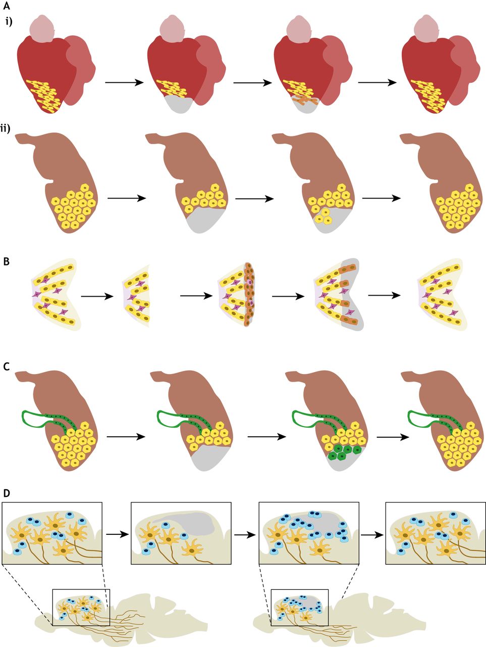
Fig. 3 Cellular origin of the de novo formed tissue during organ regeneration in the zebrafish. (A) Dedifferentiation, proliferation and re-differentiation. (Ai) In the heart, cardiomyocytes in close proximity to the injury revert to a less differentiated stage, re-enter the cell cycle and redifferentiate into mature cardiomyocytes. (Aii) During regeneration of minor liver damage, hepatocyte regeneration occurs with no signs of dedifferentiation prior to cell cycle entry and proliferation. (B) Blastema formation as an intermediate step during regeneration. After fin amputation, cells of various lineages – including osteoblasts – dedifferentiate and accumulate under an apical epidermal cap. They then proliferate and redifferentiate to rebuild the missing fin structures. (C) Phenotypic switch or transdifferentiation during regeneration. Example: after extensive liver damage, biliary ductal cells (green) can transdifferentiate into hepatocytes (green hexagonal cells) that then differentiate into mature proliferating hepatocytes. (D) Stem cells as progenitor cells. Neural stem cells/progenitor cells proliferate and differentiate into new neurons during regeneration of the central nervous system. While neuronal regeneration has been well described, less information is available on robust axon regrowth. Yellow, differentiated cells; orange, dedifferentiated cells; purple, non-osteoblast cells within the fin; green hexagonal cells, cells undergoing transdifferentiation; blue, stem cells/progenitor cells. Damaged area is shown in gray.